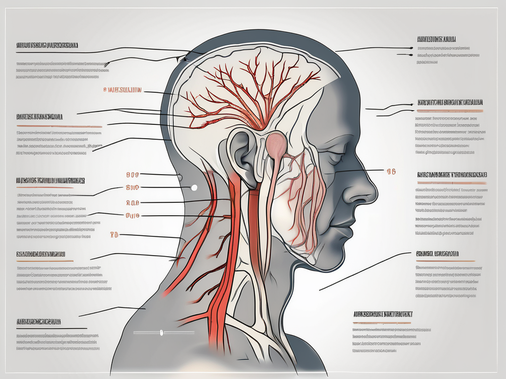
The greater auricular nerve, also known as the superficial cervical plexus, is a branch of the cervical plexus. This nerve plays a crucial role in sensory innervation of the skin on the side of the head and the external ear. Understanding the causes, symptoms, and treatment options for greater auricular nerve injury is important for both medical professionals and individuals experiencing related symptoms.
What is the Greater Auricular Nerve?
The greater auricular nerve is responsible for providing sensation to the skin around the ear, including the external ear and the area behind the earlobe. It originates from the second and third cervical nerves, known as C2 and C3, respectively. This nerve courses along the side of the neck, separated from the underlying structures by fascia and muscles.
The greater auricular nerve plays a crucial role in our everyday lives, allowing us to experience the world through touch, pain, and temperature. Without this nerve, our ability to feel the gentle breeze on our ears, the warmth of the sun, or the pain of an injury would be greatly diminished.
The Role of the Greater Auricular Nerve
The primary function of the greater auricular nerve is to transmit sensory information from the regions it innervates to the brain. By relaying signals of touch, pain, and temperature, it allows individuals to have a sense of feeling in the corresponding areas of the head and external ear.
Imagine the intricate network of nerve fibers running through your ear, constantly sending messages to your brain, allowing you to perceive the world around you. The greater auricular nerve is like a messenger, delivering these important signals and ensuring that we are aware of our surroundings.
Anatomy of the Greater Auricular Nerve
The greater auricular nerve has a complex anatomy, extending from the posterior triangle of the neck towards the ear. Along its course, it winds superficially around the sternocleidomastoid muscle. Close to the ear, it divides into branches that supply sensation to different regions around the ear and neck.
As the nerve winds its way through the neck, it is protected by layers of fascia and muscles, shielding it from potential damage. This intricate arrangement of tissues ensures that the greater auricular nerve remains safe and functional, allowing us to experience the world with clarity and sensitivity.
Understanding the anatomy of the greater auricular nerve is essential for medical professionals, as it enables them to accurately diagnose and treat conditions that may affect this important sensory pathway. By studying the intricate details of this nerve, researchers and healthcare providers can gain valuable insights into the complexities of the human body.
Causes of Greater Auricular Nerve Injury
Greater auricular nerve injury can occur due to a variety of causes, which can be classified into trauma-related, surgical, and non-traumatic causes. Understanding the different factors that can lead to this type of nerve injury is crucial for prevention and effective management.
Trauma-Related Causes
One of the primary causes of greater auricular nerve injury is trauma. Accidents, such as motor vehicle collisions, falls, or sports injuries, can result in direct trauma to the side of the head or neck. The impact from these incidents can lead to compression or stretching of the greater auricular nerve, causing damage and subsequent symptoms.
For instance, imagine a scenario where an individual is involved in a car accident. The force of the collision causes their head to forcefully strike the side window, resulting in a blow to the side of the head. This sudden impact can directly affect the greater auricular nerve, leading to injury and potential complications.
Surgical Causes
In addition to trauma, surgical procedures involving the head or neck region can also contribute to greater auricular nerve injury. Medical interventions such as facial surgeries or neck dissections, which are often performed for various reasons including cosmetic or therapeutic purposes, can inadvertently damage the greater auricular nerve.
During these surgical procedures, incisions are made and tissues are manipulated in close proximity to the nerve. While surgeons take great care to avoid nerve damage, there is always a risk of unintended injury. The delicate nature of the greater auricular nerve makes it particularly vulnerable during such interventions.
Non-Traumatic Causes
Aside from trauma and surgery, there are non-traumatic causes that can lead to greater auricular nerve injury. Infections, such as cellulitis or abscesses, can affect the nerve and result in inflammation or damage. Additionally, certain inflammatory conditions, like vasculitis or autoimmune disorders, can also impact the nerve’s function.
Furthermore, compressive neuropathies can contribute to greater auricular nerve injury. Conditions such as nerve entrapment or compression due to surrounding structures can impede the nerve’s normal function and lead to symptoms. It is important to note that underlying systemic disorders, such as diabetes, can increase the risk of nerve damage and exacerbate the effects of these non-traumatic causes.
Understanding the various causes of greater auricular nerve injury is essential for healthcare professionals to accurately diagnose and manage this condition. By identifying the underlying cause, appropriate treatment strategies can be implemented to alleviate symptoms, promote healing, and prevent further damage to the nerve.
Recognizing the Symptoms of Greater Auricular Nerve Injury
Greater auricular nerve injury can present with a range of symptoms that may vary in severity and duration.
When it comes to physical symptoms of greater auricular nerve injury, individuals may experience more than just numbness, tingling, or a burning sensation in the affected areas. In some cases, the pain can be excruciating, making it difficult for individuals to carry out their daily activities. The discomfort can radiate from the neck to the ears, causing a throbbing sensation that can be quite distressing. Furthermore, muscle weakness or atrophy in the neck can occur if the injury is severe or long-standing. This can lead to difficulties in performing tasks that require neck movement, such as turning the head or looking up and down.
In addition to physical symptoms, individuals with greater auricular nerve injury may also experience sensory symptoms. The nerve is responsible for providing sensation to certain areas of the skin, so when it is injured, individuals may report altered or decreased sensation in those areas. This can affect their ability to feel touch or changes in temperature. For example, they may have difficulty detecting whether something is hot or cold, which can be dangerous in certain situations.
Moreover, the impact of greater auricular nerve injury on daily life should not be underestimated. Depending on the extent of the injury and the individual’s occupation or lifestyle, it can have a significant effect on their day-to-day activities. For instance, wearing glasses or headphones may become a challenge due to the discomfort caused by pressure on or around the ears. This can be particularly frustrating for individuals who rely on these items for work or leisure activities. Additionally, aesthetic concerns may arise as a result of altered sensation or muscle weakness. The asymmetry in facial appearance caused by muscle atrophy can lead to self-consciousness and affect an individual’s self-esteem.
In conclusion, greater auricular nerve injury can manifest with a variety of symptoms, both physical and sensory. These symptoms can have a significant impact on an individual’s daily life, affecting their ability to perform tasks and causing aesthetic concerns. It is important to recognize these symptoms and seek appropriate medical attention to manage the injury effectively.
Diagnostic Procedures for Greater Auricular Nerve Injury
If greater auricular nerve injury is suspected, a medical professional may perform various diagnostic procedures to confirm the diagnosis and assess the severity of the injury.
Greater auricular nerve injury can occur due to various reasons, such as trauma, compression, or inflammation. The symptoms may include pain, numbness, tingling, and weakness in the areas innervated by the nerve, which include the ear, the skin over the parotid gland, and the angle of the mandible.
Clinical Examination
A clinical examination involves a thorough assessment of the affected areas, focusing on sensory deficits, muscle strength, and overall nerve function. The medical professional may gently touch different parts of the face and neck to evaluate sensation and ask the individual to perform specific movements to assess muscle function.
During the clinical examination, the medical professional may also inquire about the individual’s medical history and any previous injuries or conditions that may be contributing to the symptoms. This information helps in determining the underlying cause of the greater auricular nerve injury.
Imaging Techniques
In some cases, imaging techniques such as MRI or ultrasound may be used to visualize the anatomy of the greater auricular nerve or identify any structural abnormalities or lesions that could be causing the symptoms.
Magnetic Resonance Imaging (MRI) provides detailed images of the soft tissues and can help identify any nerve compression or inflammation. Ultrasound, on the other hand, uses sound waves to create real-time images and can be useful in assessing the nerve’s structure and any potential abnormalities.
Electrophysiological Studies
Electrophysiological studies, such as nerve conduction studies or electromyography, may be performed to assess the conduction of nerve signals and detect any abnormalities in the greater auricular nerve.
Nerve conduction studies involve the application of small electrical impulses to the nerve and measuring the response. This helps in evaluating the nerve’s ability to transmit signals and can identify any areas of damage or dysfunction.
Electromyography (EMG) involves the insertion of tiny needles into the muscles innervated by the greater auricular nerve. The electrical activity of the muscles is then recorded, providing information about the nerve-muscle connection and any potential abnormalities.
It is important to note that self-diagnosis is not recommended, and individuals experiencing symptoms of greater auricular nerve injury should consult a medical professional for proper evaluation and diagnosis.
A timely and accurate diagnosis is crucial for determining the appropriate treatment plan and preventing further complications. With the help of diagnostic procedures, medical professionals can gather valuable information about the extent and underlying cause of the greater auricular nerve injury, leading to effective management and rehabilitation strategies.
Leave a Reply