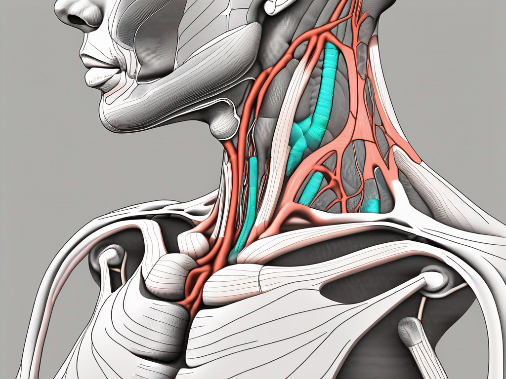
The great auricular nerve is an essential component of the human nervous system, responsible for transmitting sensory information from the skin of the ear and the region behind it. Its anatomical relationship with the sternocleidomastoid muscle is of particular interest and clinical significance. Understanding the intricate interplay between these structures can provide valuable insights into various health issues and guide medical interventions when necessary.
Understanding the Anatomy of the Great Auricular Nerve
The great auricular nerve is a fascinating component of the human body’s intricate nervous system. Originating from the cervical plexus, specifically from the spinal nerves C2 and C3, this nerve serves a crucial role in providing sensory feedback to various regions of the head and neck.
As its name suggests, the great auricular nerve primarily innervates the skin overlying the external ear. However, its reach extends beyond this area, also supplying sensation to parts of the parotid gland. This nerve’s ability to transmit sensory information is essential for our perception of temperature, touch, and pain in these regions.
Origin and Pathway of the Great Auricular Nerve
The journey of the great auricular nerve begins at the cervical plexus, where it arises from the anterior rami of the second and third cervical spinal nerves, known as C2 and C3, respectively. From its origin, this nerve emerges near the posterior border of the sternocleidomastoid muscle.
Traversing across the sternocleidomastoid muscle, the great auricular nerve follows a distinct pathway towards its destination. Along this pathway, it branches out, sending sensory fibers to the skin overlying the external ear and the areas extending behind it. This intricate pathway ensures efficient transmission of sensory information, allowing us to perceive various sensations in these regions.
Function and Clinical Significance of the Great Auricular Nerve
The great auricular nerve’s primary function is to provide sensory innervation to the skin overlying the external ear and the regions extending behind it. Through its sensory fibers, this nerve enables us to perceive the touch of a gentle breeze, the warmth of the sun, and the pain from an accidental bump.
Understanding the clinical significance of the great auricular nerve is crucial for diagnosing and managing related health issues effectively. In certain cases, this nerve can be subject to entrapment or develop lesions, leading to sensory disturbances and discomfort. Recognizing these clinical conditions and their impact on the great auricular nerve is essential for providing appropriate medical interventions and alleviating patients’ symptoms.
The great auricular nerve’s intricate anatomy and vital role in sensory perception highlight its significance in the human body. By expanding our knowledge of this nerve’s origin, pathway, and function, we gain a deeper understanding of the complexity and interconnectedness of our physiological systems.
The Sternocleidomastoid Muscle: An Overview
The sternocleidomastoid muscle is one of the most prominent structures in the anterior neck region. Its location, structure, and physiological functions contribute to its significance in various aspects of human anatomy and movement.
Structure and Location of the Sternocleidomastoid Muscle
The sternocleidomastoid muscle is a paired muscle that originates from the sternum and clavicle and inserts onto the mastoid process of the temporal bone. Its positioning creates a diagonal line across the anterior neck region. This muscle is easily palpable and visible in individuals, serving as an important landmark for medical professionals during physical examinations and surgical procedures.
When examining the structure of the sternocleidomastoid muscle, it is important to note that it is divided into two heads: the sternal head and the clavicular head. The sternal head originates from the manubrium of the sternum, while the clavicular head originates from the medial third of the clavicle. These two heads merge together to form a thick muscle belly, which then tapers and inserts onto the mastoid process of the temporal bone.
As for the location of the sternocleidomastoid muscle, it is situated superficially in the anterior neck region. It spans from the sternum and clavicle to the mastoid process, creating a visible and palpable prominence. Its diagonal orientation across the neck gives it a distinct appearance, making it easily identifiable.
Role and Importance of the Sternocleidomastoid Muscle in the Human Body
The sternocleidomastoid muscle performs several crucial functions. It allows rotation and lateral flexion of the head and neck. This means that it enables us to turn our heads from side to side and tilt our heads towards our shoulders. These movements are essential for various activities in our daily lives, such as looking over our shoulders while driving, turning our heads to follow a conversation, or tilting our heads to rest on our shoulders when we are tired.
In addition to its role in head movements, the sternocleidomastoid muscle also plays a significant role in maintaining proper posture. It works in conjunction with other muscles in the neck and upper back to keep the head aligned with the spine. This helps prevent slouching and promotes an upright posture, which is important for overall musculoskeletal health.
Furthermore, the sternocleidomastoid muscle acts as a stabilizer for the head during various activities. Whether we are running, jumping, or engaging in any physical movement, this muscle helps keep our head steady and prevents excessive movement that could lead to injury. It provides stability and control, allowing us to perform tasks with precision and accuracy.
It is worth noting that dysfunction or injury to the sternocleidomastoid muscle can result in limitations in head movements and contribute to neck pain. Conditions such as muscle strains, trigger points, or even congenital torticollis can affect the function of this muscle. Therefore, understanding the role and importance of the sternocleidomastoid muscle is paramount for healthcare practitioners when evaluating patients with related symptoms.
The Intersection of the Great Auricular Nerve and Sternocleidomastoid Muscle
The area where the great auricular nerve intersects with the sternocleidomastoid muscle is of particular interest due to its clinical implications. The anatomical relationship between these structures can significantly impact medical procedures and diagnostic protocols.
The Path of the Great Auricular Nerve across the Sternocleidomastoid
As the great auricular nerve emerges near the posterior border of the sternocleidomastoid muscle, it courses obliquely across its surface. This path exposes the nerve to potential compression or injury, especially during surgical interventions or certain medical procedures. Knowledge of this course is essential to minimize the risk of damage to the great auricular nerve and facilitate optimal outcomes.
Implications of the Intersection on Medical Procedures
Medical procedures involving the sternocleidomastoid region, such as surgeries, injections, or nerve blocks, should consider the presence and vulnerability of the great auricular nerve. Careful attention should be given to its location and pathway to avoid iatrogenic nerve injury or unintended sensory deficits. Consulting with a healthcare professional experienced in neuroanatomy and the specific procedure is crucial to ensure the best possible outcome.
Potential Health Issues Related to the Great Auricular Nerve and Sternocleidomastoid Muscle
Several health issues can arise in association with the great auricular nerve and the sternocleidomastoid muscle. Understanding these conditions, their symptoms, and available diagnostic and treatment options is imperative for comprehensive patient care.
Common Disorders and Their Symptoms
Disorders affecting the great auricular nerve and the sternocleidomastoid muscle can manifest in various ways. Patients may experience pain, altered sensation, or even motor deficits in the associated areas. Conditions such as nerve entrapment, inflammation, or trauma can affect the functionality of these structures, potentially leading to discomfort and functional limitations. If you experience any symptoms related to the ear, neck, or surrounding regions, it is advisable to consult with a healthcare professional for accurate diagnosis and appropriate management options.
Diagnostic Methods and Treatment Options
The diagnosis of health issues involving the great auricular nerve and sternocleidomastoid muscle often relies on a thorough physical examination, clinical assessment, and possibly additional imaging studies. Treatment strategies may range from conservative approaches, including medications and physical therapy, to more invasive interventions like nerve blocks or surgical procedures. In all cases, the expertise of a healthcare provider familiar with the specific condition is indispensable, as individual cases may vary, and personalized management plans should be tailored accordingly.
Future Research Directions in Neuroanatomy
Advancements in technology and the continuous pursuit of knowledge have opened up exciting avenues for further exploration in the field of neuroanatomy. Several areas related to the great auricular nerve and sternocleidomastoid muscle warrant further investigation to enhance our understanding and improve clinical practices.
Unanswered Questions about the Great Auricular Nerve and Sternocleidomastoid Muscle
Despite significant progress in neuroanatomy research, there are still unanswered questions regarding the great auricular nerve and its relationship with the sternocleidomastoid muscle. The precise mechanisms of nerve function, potential variations in anatomy, and the effects of aging or pathological conditions all warrant further investigation for a more comprehensive understanding.
The Impact of Technological Advancements on Neuroanatomy Research
The development of advanced imaging techniques, such as magnetic resonance imaging (MRI) and ultrasound, has revolutionized neuroanatomy research. These technologies allow for non-invasive visualization and detailed analysis of nerve structures, providing valuable insights into their relationships and functions. Continued technological advancements will likely drive further breakthroughs, expanding our understanding of the great auricular nerve, the sternocleidomastoid muscle, and their intricate interactions.
In conclusion, the intersection of the great auricular nerve and the posterior border of the sternocleidomastoid muscle is an anatomical site of paramount importance. Knowledge of the anatomy, function, and clinical significance of these structures is crucial for healthcare professionals involved in the diagnosis and treatment of related health issues. As research continues to evolve, these insights will pave the way for improved patient care and surgical interventions, ensuring optimal outcomes and minimizing risks. If you have any concerns or experience symptoms related to the areas discussed in this article, it is always advised to consult with your healthcare provider for appropriate evaluation and guidance.
Leave a Reply