The greater auricular nerve is a crucial component of the human nervous system, playing a vital role as a general sensory afferent. Its intricate anatomy and physiological functions have intrigued researchers and healthcare professionals alike. By delving into its various aspects, we can gain a deeper understanding of its significance in medical practice and potential therapeutic applications. This article aims to explore the anatomy, mechanism of sensory transmission, clinical significance, and current research surrounding the greater auricular nerve.
Anatomy of the Greater Auricular Nerve
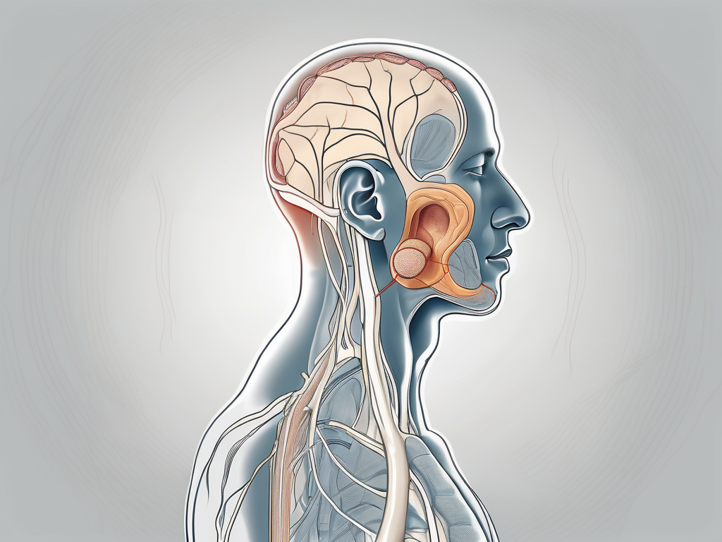
The greater auricular nerve is a branch of the cervical plexus, arising from the second and third cervical spinal nerves (C2-C3) in the neck region. It courses superficially over the sternocleidomastoid muscle, extending towards the ear.
The greater auricular nerve, also known as the auricular branch of the cervical plexus, plays a crucial role in the sensory innervation of the head and neck region. It is responsible for transmitting sensory information from specific areas to the brain, allowing us to perceive touch, temperature, and pain.
As it courses superficially over the sternocleidomastoid muscle, the greater auricular nerve branches out and distributes its sensory fibers to various regions of the head and neck. One of its primary areas of innervation is the skin over the parotid gland, which is located in front of the ear. This region includes the cheek and the area just below the earlobe.
In addition to the parotid gland, the greater auricular nerve also innervates the external ear. It provides sensory information from the skin of the ear, allowing us to perceive touch and temperature. This is particularly important for our ability to hear, as the external ear plays a crucial role in capturing sound waves and transmitting them to the middle ear.
Furthermore, the greater auricular nerve extends its branches to the mastoid process, a prominent bony projection located behind the ear. The mastoid process is an important anatomical landmark and serves as an attachment site for several muscles involved in head and neck movements. The sensory innervation provided by the greater auricular nerve to this region allows us to perceive touch and pressure, contributing to our overall spatial awareness.
In summary, the greater auricular nerve is a vital component of the cervical plexus, responsible for the sensory innervation of the head and neck region. Its branches distribute sensory fibers to the skin over the parotid gland, external ear, and mastoid process, allowing us to perceive touch, temperature, and pain in these areas.
The Greater Auricular Nerve as a Sensory Afferent
Understanding the concept of sensory afferents is crucial to comprehending the role of the greater auricular nerve. Sensory afferents are nerve fibers that transmit sensory information from the peripheral tissues to the central nervous system. They play a pivotal role in our perception and response to various stimuli.
The greater auricular nerve, a branch of the cervical plexus, is one such sensory afferent that holds significant importance in the realm of sensation. This nerve is responsible for innervating the skin overlying the external ear and a portion of the parotid gland. It carries vital sensory information from these areas to the brain, allowing us to perceive touch, temperature, and pain in this region.
Mechanism of Sensory Transmission in the Greater Auricular Nerve
When stimulated by touch, temperature, or pain, specialized receptors in the skin generate electrical signals. These signals are then transmitted along the greater auricular nerve fibers through a process called action potential. The nerve fibers act as conduits, relaying the information to the brain for interpretation and response.
Let’s delve deeper into the fascinating process of sensory transmission in the greater auricular nerve. When an external stimulus, such as a gentle touch on the earlobe, is applied, mechanoreceptors in the skin detect the pressure and convert it into electrical signals. These mechanoreceptors, known as Merkel cells and Meissner’s corpuscles, are highly sensitive to changes in pressure and play a crucial role in our ability to perceive tactile sensations.
Once the electrical signals are generated, they travel along the nerve fibers of the greater auricular nerve. These nerve fibers are composed of individual cells called neurons, which are specialized in transmitting electrical impulses. The transmission of these signals occurs through a series of intricate processes within the nerve fibers.
Firstly, the electrical signals trigger the opening of ion channels in the cell membrane of the neurons. This allows the influx of positively charged ions, such as sodium and potassium, into the neuron, leading to a change in its electrical potential. This change, known as depolarization, propagates along the nerve fiber, creating a wave-like action potential.
As the action potential travels along the nerve fiber, it triggers the opening of voltage-gated ion channels at specific intervals. These channels ensure that the action potential remains strong and does not diminish as it travels towards the brain. The opening and closing of these ion channels create a domino effect, ensuring the efficient transmission of the electrical signals.
Once the action potential reaches the end of the greater auricular nerve fibers, it encounters specialized structures called synapses. Synapses are junctions between two neurons, where the electrical signals are converted into chemical signals for transmission to the next neuron. In the case of sensory transmission in the greater auricular nerve, the synapses connect the nerve fibers to neurons in the brainstem and thalamus.
At the synapses, the electrical signals trigger the release of neurotransmitters, which are chemical messengers that allow communication between neurons. These neurotransmitters diffuse across the synapse and bind to receptors on the receiving neuron, initiating a new electrical signal. This signal then continues its journey through the intricate network of neurons in the central nervous system, ultimately reaching the brain for interpretation and response.
The process of sensory transmission in the greater auricular nerve is a remarkable feat of biological engineering. It allows us to experience the world around us, perceive sensations, and respond accordingly. From the gentle touch of a loved one’s hand on our ear to the warning signal of a hot object, the greater auricular nerve plays a vital role in our daily lives, ensuring our safety and enhancing our sensory experiences.
Clinical Significance of the Greater Auricular Nerve
The greater auricular nerve holds significant clinical relevance, particularly in surgical procedures and the diagnosis of neurological disorders.
The greater auricular nerve, also known as the auricular branch of the cervical plexus, is a sensory nerve that arises from the cervical spinal nerves C2 and C3. It provides innervation to the skin over the external ear, the parotid gland, and the angle of the mandible.
Role in Surgical Procedures
During certain surgical interventions, the greater auricular nerve can be used as a reliable anatomical landmark. It assists surgeons in identifying critical structures and performing precise surgical techniques.
For example, in plastic and reconstructive surgery, the greater auricular nerve can be utilized for nerve grafting procedures. Nerve grafting involves taking a segment of the greater auricular nerve and using it to repair damaged or severed nerves in other parts of the body, restoring sensory and motor function.
Furthermore, the greater auricular nerve can be used as a guide during surgical procedures involving the parotid gland, such as parotidectomy or facial nerve decompression. Its close proximity to the gland allows surgeons to locate and protect the nerve while removing tumors or addressing other pathologies.
However, it’s crucial to note that any surgical procedure involving the greater auricular nerve should be conducted by qualified healthcare professionals. These professionals have the necessary expertise to minimize the risks and ensure optimal outcomes.
If you are considering a surgical procedure that involves the greater auricular nerve, it is essential to schedule a consultation with a specialist. During the consultation, you can discuss your specific concerns and the potential benefits and risks associated with the procedure. The specialist will provide you with personalized advice and guidance, taking into account your unique medical history and circumstances.
Implications in Neurological Disorders
The greater auricular nerve’s involvement in neurological disorders warrants attention. Pathologies affecting the nerve, such as trauma or compression, can result in altered sensory perception, pain, or even sensory loss in the innervated regions.
For instance, trauma to the greater auricular nerve, such as a direct injury or surgical manipulation, can lead to neuropathic pain or paresthesia in the area supplied by the nerve. This can significantly impact a person’s quality of life and may require specialized treatment approaches, such as nerve blocks or medications targeting neuropathic pain.
In addition to trauma, compression of the greater auricular nerve can occur due to various factors, including tight headwear, prolonged pressure, or certain medical conditions. This compression can result in symptoms such as numbness, tingling, or a burning sensation in the distribution of the nerve.
If you experience any unusual symptoms or changes in sensation in the areas supplied by the greater auricular nerve, it is crucial to consult a healthcare provider. They will be able to evaluate your symptoms, perform a thorough examination, and order any necessary diagnostic tests to determine the underlying cause. Early diagnosis and appropriate medical intervention can help manage and potentially reverse the effects of neurological disorders affecting the greater auricular nerve.
In conclusion, the greater auricular nerve plays a vital role in both surgical procedures and the diagnosis of neurological disorders. Its anatomical significance and potential implications highlight the importance of understanding its function and seeking professional medical advice when necessary.
Research and Future Directions
Continued research on the greater auricular nerve sheds light on its complex functions and potential therapeutic applications.
Current Research on the Greater Auricular Nerve
Researchers are actively investigating various aspects of the greater auricular nerve, including its physiological properties, molecular pathways, and clinical implications. These studies aim to enhance our understanding of the nerve’s role and uncover novel ways to diagnose and treat related conditions.
Potential Therapeutic Applications and Future Research Directions
In the future, harnessing the knowledge gained from current research may open doors to innovative therapeutic approaches. For instance, nerve stimulation techniques targeting the greater auricular nerve hold promise in managing chronic pain and neurological disorders. However, it is essential to consult a healthcare professional to discuss the suitability and safety of such approaches.
In conclusion, the greater auricular nerve serves as a vital general sensory afferent, enabling us to perceive touch, temperature, and pain in specific regions of the head and neck. Through understanding its anatomy, mechanism of transmission, clinical significance, and ongoing research, we can appreciate the integral role this nerve plays in our overall sensory experience. Should you have any concerns or questions related to the greater auricular nerve, seek guidance from a qualified healthcare professional.
Leave a Reply