In this comprehensive guide, we will delve into the intricate world of the Great Auricular Nerve and the Auriculotemporal Nerve. These nerves play crucial roles in the human body, and understanding their anatomy, functions, clinical significance, and associated disorders is essential for anyone seeking a holistic understanding of the nervous system. So, let’s embark on this journey and explore the fascinating realm of these nerves.
Anatomy of the Great Auricular Nerve
The Great Auricular Nerve is a significant nerve that plays a crucial role in providing sensory innervation to specific areas of the head and neck. Let’s take a closer look at its origin, pathway, functions, and clinical significance.
Origin and Pathway of the Great Auricular Nerve
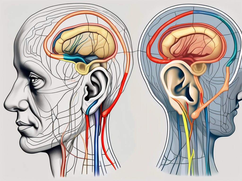
The Great Auricular Nerve originates from the cervical plexus, which is a network of nerves formed by the anterior rami of the spinal nerves C2 and C3. From its origin, it emerges from the posterior border of the sternocleidomastoid muscle, a large muscle located in the neck. As it ascends obliquely, it closely follows the posterior border of the external jugular vein, a major vein in the neck. Continuing on its pathway, the nerve travels towards the ear and the angle of the mandible, providing sensory innervation along its course.
One of the primary areas innervated by the Great Auricular Nerve is the skin over the parotid gland, which is a salivary gland located in front of the ear. Additionally, it supplies sensory innervation to the external ear and the mastoid process, a prominent bony projection located behind the ear. This extensive innervation allows for the transmission of various sensations from these areas to the brain.
Functions of the Great Auricular Nerve
The Great Auricular Nerve primarily functions as a sensory nerve, transmitting important sensations such as pain, temperature, and touch from the designated areas to the brain. This sensory information is crucial for our perception of the external environment and our ability to respond to stimuli appropriately.
In addition to its sensory role, the Great Auricular Nerve also contributes to the motor innervation of the platysma muscle. The platysma muscle is a thin, broad muscle that covers the front of the neck. The involvement of the Great Auricular Nerve in the motor innervation of this muscle allows for certain movements of the neck, such as tilting the head or turning it to the side.
Clinical Significance of the Great Auricular Nerve
Understanding the clinical significance of the Great Auricular Nerve is vital, especially in the context of certain medical procedures. Surgeons performing surgeries such as parotid gland removal or facelifts must carefully identify and preserve this nerve to avoid potential complications.
Damage to the Great Auricular Nerve during surgical procedures can result in the loss of sensory function in the areas it innervates. This loss of sensation can have a significant impact on a patient’s quality of life, affecting their ability to perceive touch, temperature, and pain in the affected areas.
In some cases, injury or compression of the Great Auricular Nerve can occur independently of surgical procedures. This can happen due to trauma, such as a direct blow to the neck or head, or due to conditions that cause nerve compression, such as tumors or inflammation. When the nerve is injured or compressed, it can lead to symptoms such as pain, altered sensation, or even numbness in the areas it supplies. Prompt medical attention is necessary in such cases to diagnose and manage the underlying cause and prevent any long-term complications.
In conclusion, the Great Auricular Nerve is an essential nerve that provides sensory innervation to specific areas of the head and neck. Its origin, pathway, functions, and clinical significance highlight its importance in maintaining normal sensation and motor function in these regions.
Anatomy of the Auriculotemporal Nerve
The Auriculotemporal Nerve is a significant nerve that plays a crucial role in the sensory innervation of the head and face. Let’s explore its origin, pathway, functions, and clinical significance in more detail.
Origin and Pathway of the Auriculotemporal Nerve
The Auriculotemporal Nerve arises from the mandibular division of the trigeminal nerve (V3), which is one of the twelve cranial nerves. This division carries sensory information from the lower face, jaw, and parts of the scalp. As the Auriculotemporal Nerve emerges from the mandibular division, it travels posteriorly from the infratemporal fossa and enters the parotid gland, a large salivary gland located in front of the ear.
Within the parotid gland, the Auriculotemporal Nerve branches out into smaller nerve fibers, which then provide sensory innervation to specific areas. These areas include the external ear, the skin covering the temporal region (the sides of the head), and the scalp behind the ear. The nerve fibers extend throughout these regions, allowing for the transmission of sensory information to the brain.
Functions of the Auriculotemporal Nerve
The primary function of the Auriculotemporal Nerve is to carry sensory information from the designated areas to the brain. This nerve enables the perception of pain, temperature, and touch, allowing us to experience and respond to various stimuli accordingly. For example, when we feel a gentle breeze on our scalp or experience a sharp pain in our ear, it is the Auriculotemporal Nerve that conveys these sensations to the brain for interpretation.
In addition to its sensory role, the Auriculotemporal Nerve also contributes to the autonomic innervation of the parotid gland. This means that it plays a vital role in regulating the gland’s function, particularly in salivation. The nerve fibers within the Auriculotemporal Nerve carry signals that control the production and release of saliva, ensuring proper oral health and digestion.
Clinical Significance of the Auriculotemporal Nerve
The Auriculotemporal Nerve holds significant clinical importance, especially in certain medical procedures. For instance, during dental work or temporomandibular joint (TMJ) surgery, there is a potential risk of affecting the Auriculotemporal Nerve. Injury to this nerve can lead to diminished sensation in the areas it serves, resulting in discomfort and possible functional disturbances.
Therefore, it is crucial for healthcare professionals to approach these procedures with caution, taking into account the delicacy and importance of the Auriculotemporal Nerve. Careful planning and precise execution can help minimize the risk of nerve injury and ensure optimal patient outcomes.
In conclusion, the Auriculotemporal Nerve is a vital component of the trigeminal nerve and plays a significant role in the sensory innervation of the head and face. Understanding its anatomy, functions, and clinical significance is essential for healthcare professionals involved in procedures that may potentially affect this nerve. By prioritizing its preservation and function, we can ensure the overall well-being and comfort of patients.
Understanding the Nervous System
Role of Nerves in the Human Body
The nervous system is a complex network that regulates and coordinates various bodily functions. Nerves, including the Great Auricular Nerve and the Auriculotemporal Nerve, serve as the messengers, transmitting signals between different parts of the body and the brain. They allow us to perceive our surroundings, experience sensations, and initiate appropriate responses.
How Nerves Communicate with the Brain
Nerves communicate with the brain through a process called neurotransmission. When stimulated, nerves generate electrical impulses that travel along their length. At the ends of the nerves, near the synapses, these electrical signals are converted into chemical messages (neurotransmitters) that cross the synapse and transmit the information to the next nerve or target organ. This intricate communication system enables the brain to receive and interpret signals from various parts of the body.
Disorders Related to the Great Auricular and Auriculotemporal Nerves
Symptoms and Diagnosis
Disorders related to the Great Auricular Nerve and the Auriculotemporal Nerve can manifest in various ways. Common symptoms may include pain, altered sensation, numbness, or tingling in the designated areas. Diagnosing these disorders often involves a detailed medical history, physical examination, and sometimes imaging studies or nerve conduction tests.
Treatment Options and Recovery
The treatment of these nerve-related disorders depends on the underlying cause and the severity of the symptoms. It is essential to consult with a healthcare professional for an accurate diagnosis and appropriate treatment options. Treatment may include medications for pain management, physical therapy, lifestyle modifications, and in some cases, surgical interventions. Recovery time varies, and it is crucial to follow the prescribed treatment plan and allow the body sufficient time to heal.
Prevention and Management of Nerve Disorders
Lifestyle Changes for Nerve Health
Maintaining good overall health can contribute to optimal nerve function. Engaging in regular exercise, following a balanced diet, managing stress levels, and avoiding lifestyle factors that may harm nerves are essential. It is also crucial to protect yourself from injury, as trauma can potentially affect the function of the Great Auricular Nerve and the Auriculotemporal Nerve.
Medical Interventions for Nerve Protection
In certain cases, medical interventions may be necessary to protect and support nerve function. This may include the use of appropriate protective equipment during high-risk activities, such as sports or certain occupations. Additionally, managing underlying medical conditions that may affect nerve health, such as diabetes or autoimmune disorders, is crucial in preventing potential nerve damage. Seeking guidance from healthcare professionals will help ensure the best course of action.
In conclusion, understanding the Great Auricular Nerve and the Auriculotemporal Nerve is a crucial step towards comprehending the complexity of the nervous system. These nerves play vital roles in transmitting sensory information and contributing to motor function. Recognizing their anatomy, functions, clinical significance, and potential disorders is essential for healthcare professionals and individuals seeking profound knowledge in the realm of the nervous system. Remember, if you experience any symptoms or concerns related to these nerves, consult with a medical professional to receive appropriate guidance and care.
Leave a Reply