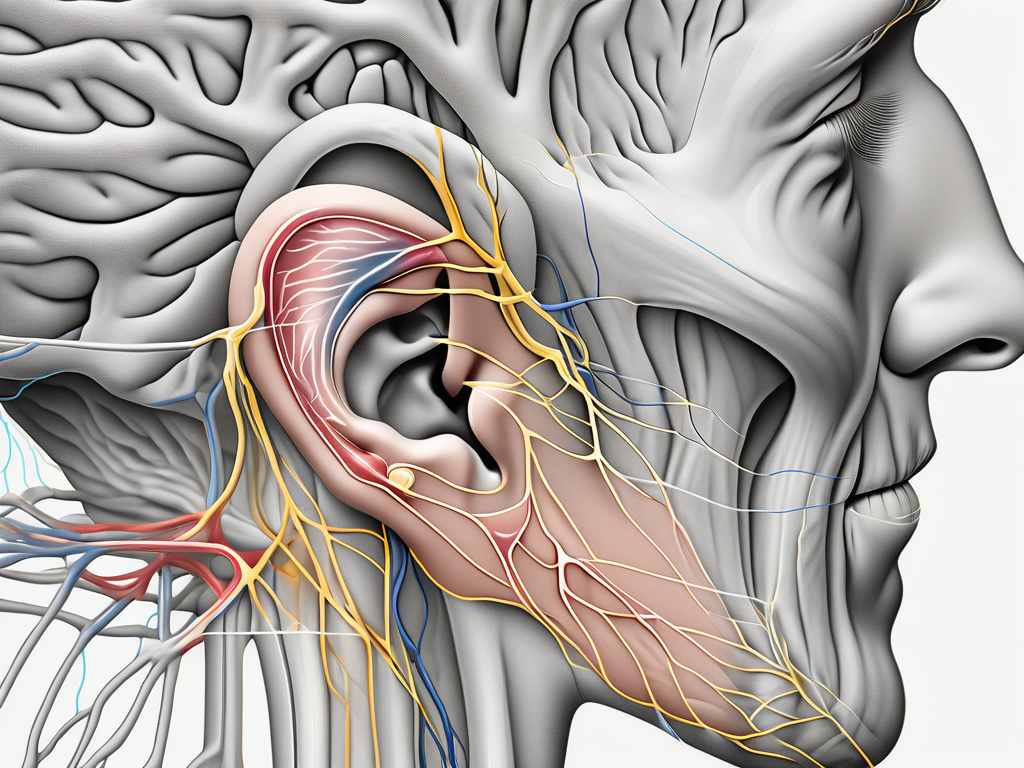
The posterior auricular nerve is a crucial component of the peripheral nervous system, playing a vital role in the functioning of the human body. Its distribution and function have intrigued medical experts for years, and through countless research and discoveries, our understanding of this nerve has significantly improved. In this article, we will explore the anatomy, functions, clinical significance, and recent developments surrounding the posterior auricular nerve.
Anatomy of the Posterior Auricular Nerve
The posterior auricular nerve is a branch of the facial nerve, specifically emerging from the stylomastoid foramen. This foramen serves as the exit point for various nerves involved in facial sensation and movement. Upon leaving the stylomastoid foramen, the posterior auricular nerve travels posteriorly along the mastoid process of the temporal bone.
The posterior auricular nerve plays a crucial role in the sensory innervation of the ear and surrounding areas. Let’s explore its origin, pathway, branches, and connections in more detail.
Origin and Pathway of the Posterior Auricular Nerve
Originating from the facial nerve, the posterior auricular nerve courses upwards, traversing through the parotid gland. This gland, located in front of the ear, is responsible for producing saliva and is also involved in the process of chewing. As the posterior auricular nerve passes through the parotid gland, it continues its journey towards the ear.
As the nerve approaches the ear, it divides into multiple smaller nerves, each with its own unique function and destination. These smaller nerves innervate different structures within the region of the ear, ensuring proper sensory perception and motor control.
Branches and Connections of the Posterior Auricular Nerve
The posterior auricular nerve has several branches and connections that contribute to its intricate network. One notable branch is the auricular branch, which supplies sensory innervation to the posterior part of the external ear and the skin behind the ear. This branch is responsible for transmitting sensations such as touch, temperature, and pain from these areas to the brain.
In addition to its branches, the posterior auricular nerve establishes connections with other nerves in the area, forming a complex sensory network. These connections allow for the integration of sensory information from various sources, ensuring accurate perception and response to stimuli.
Furthermore, the posterior auricular nerve interacts with neighboring structures, such as blood vessels and lymph nodes, as it courses through the ear region. These interactions play a role in maintaining the overall health and function of the area.
Understanding the anatomy of the posterior auricular nerve is essential for healthcare professionals, as it helps in diagnosing and treating conditions related to the ear and surrounding areas. By comprehending the intricate pathways and connections of this nerve, medical practitioners can provide targeted interventions to alleviate pain, restore function, and improve the overall well-being of their patients.
Functions of the Posterior Auricular Nerve
The posterior auricular nerve serves various important functions within the human body. Let’s delve into its role in sensory perception and motor control.
The posterior auricular nerve, also known as the auricular branch of the facial nerve, is a small nerve that originates from the facial nerve. It travels behind the ear and branches out to provide sensory and motor innervation to specific areas.
Role in Sensory Perception
The sensory branches of the posterior auricular nerve play a vital role in providing sensation to the skin behind the ear and the posterior part of the external ear. This allows us to perceive touch, pain, and temperature in these areas, contributing to our overall sensory experience.
When the posterior auricular nerve is stimulated by touch, specialized nerve endings called mechanoreceptors detect the pressure and send signals to the brain. These signals are then interpreted, allowing us to feel the sensation of touch behind the ear. Similarly, when exposed to extreme temperatures or injured, the nerve endings in the posterior auricular region transmit signals of pain to the brain, alerting us to potential harm or injury.
Furthermore, the posterior auricular nerve also contains thermoreceptors, which are responsible for detecting changes in temperature. This enables us to perceive the warmth of sunlight or the coolness of a breeze against the skin behind the ear.
Contribution to Motor Control
In addition to its sensory functions, the posterior auricular nerve also contributes to motor control. Through its connections within the facial nerve, it participates in the complex coordination of facial muscles, allowing for facial expressions, such as raising the eyebrows and wrinkling the forehead.
The facial nerve, which is responsible for the motor control of the muscles of facial expression, sends branches to the posterior auricular nerve. These branches carry signals from the brain to the muscles behind the ear, allowing for precise control and movement. When we raise our eyebrows in surprise or furrow our forehead in concentration, the posterior auricular nerve plays a crucial role in executing these facial expressions.
Moreover, the posterior auricular nerve also contributes to the movement of the ear itself. It innervates certain muscles that are responsible for subtle movements of the external ear, such as rotating or twitching. These movements help us in localizing sounds and enhancing our hearing capabilities.
In conclusion, the posterior auricular nerve is not only involved in sensory perception, allowing us to feel touch and temperature behind the ear, but it also plays a significant role in motor control, enabling facial expressions and subtle movements of the external ear. Its intricate connections within the facial nerve highlight its importance in the overall functioning of the human body.
Clinical Significance of the Posterior Auricular Nerve
The posterior auricular nerve, also known as the great auricular nerve, plays a crucial role in the sensory innervation of the external ear and the surrounding area. It is a branch of the facial nerve, specifically the facial nerve’s posterior auricular branch.
This nerve has significant clinical implications in terms of disorders, symptoms, and diagnostic techniques. Understanding its importance can help healthcare professionals accurately diagnose and manage conditions that affect this nerve.
Common Disorders and Symptoms
While disorders specifically affecting the posterior auricular nerve are relatively uncommon, they can occur. One such disorder is Bell’s palsy, a condition characterized by partial or complete facial paralysis. In some cases, the posterior auricular nerve can be affected, leading to symptoms such as drooping of the earlobe or difficulty moving the ear.
Another condition associated with the posterior auricular nerve is trigeminal neuralgia. This condition causes severe facial pain, often triggered by simple activities such as eating or talking. While trigeminal neuralgia primarily affects the trigeminal nerve, the posterior auricular nerve can also be involved, leading to additional pain and discomfort in the ear region.
If you experience any unusual sensations, pain, or facial weakness, it is important to consult with a healthcare professional for proper evaluation and guidance. They can perform a thorough examination and determine if the posterior auricular nerve is involved in your symptoms.
Diagnostic Techniques and Treatment Options
Medical practitioners utilize various diagnostic techniques to assess the functionality of the posterior auricular nerve and identify potential abnormalities. One commonly used method is a physical examination, where the healthcare professional evaluates the patient’s ability to move the ear, tests for sensitivity in the ear region, and checks for any visible abnormalities.
In some cases, imaging studies such as magnetic resonance imaging (MRI) or computed tomography (CT) scans may be ordered to obtain a detailed view of the nerve and surrounding structures. These imaging techniques can help identify any structural abnormalities or nerve compression that may be affecting the posterior auricular nerve.
Electrodiagnostic tests, such as nerve conduction studies or electromyography, may also be performed to assess the nerve’s electrical activity and determine if there is any nerve damage or dysfunction.
Treatment options for disorders that affect the posterior auricular nerve may vary depending on the underlying cause and severity. In cases of Bell’s palsy, the primary focus is often on managing symptoms and promoting nerve healing. This can involve medications, physical therapy, and sometimes surgical interventions.
For trigeminal neuralgia, treatment options may include medications to manage pain, nerve blocks to provide temporary relief, or surgical procedures to alleviate nerve compression and reduce pain signals.
It is imperative to consult with a qualified healthcare professional for accurate diagnosis and appropriate treatment options. They can assess your specific condition, consider your medical history, and develop a personalized treatment plan to address the disorder affecting the posterior auricular nerve.
Recent Research and Discoveries
Advances in Neurological Understanding
Ongoing research into the posterior auricular nerve has led to significant advancements in our understanding of its intricate pathways, connections, and overall functioning. These findings have shed light on the complex interplay between the peripheral nervous system and other structures in the head and neck region, further deepening our knowledge of neurological processes.
Implications for Future Medical Practice
The evolving understanding of the posterior auricular nerve holds potential implications for future medical practice. This increased knowledge may lead to advancements in diagnostic techniques, treatment modalities, and the development of more targeted therapeutic interventions. By staying at the forefront of research and embracing technological advancements, healthcare professionals can enhance patient care and improve outcomes.
In conclusion, the posterior auricular nerve plays a critical role in our sensory perception and motor control. Understanding its distribution, anatomy, functions, and clinical significance contributes to the broader field of neurology and helps healthcare professionals provide informed care. As medical research continues to expand our knowledge, further discoveries will undoubtedly enhance our comprehension of the intricate workings of the posterior auricular nerve, ultimately benefiting patient care and advancing medical practice.

Leave a Reply