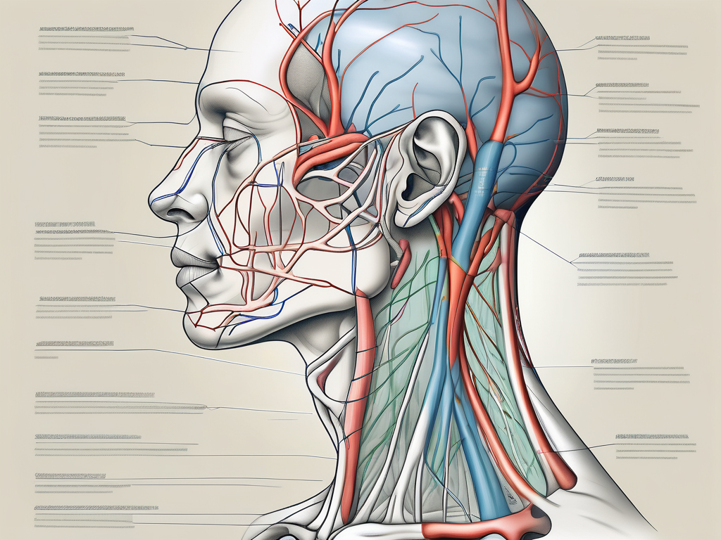
The external jugular vein and great auricular nerve are two anatomical structures that are closely related and play important roles in the human body. Understanding their anatomy, functions, and the connection between them is crucial for medical professionals and researchers in various fields. In this article, we will explore the relationship between the external jugular vein and great auricular nerve, including their individual characteristics and how they interact with each other.
Understanding the External Jugular Vein
The external jugular vein is a major blood vessel located in the neck region. It plays a vital role in the circulatory system, serving as a pathway for deoxygenated blood to return to the heart. Understanding the anatomy and functions of the external jugular vein is crucial for medical professionals and anyone interested in human physiology.
Anatomy of the External Jugular Vein
The external jugular vein is formed by the convergence of the posterior division of the retromandibular vein and the posterior auricular vein. These veins come together to create a larger vessel that runs superficially along the lateral side of the neck, just below the skin. This positioning makes the external jugular vein easily accessible for medical procedures and examinations.
When examining the external jugular vein, it is important to note its close proximity to other structures in the neck region. One such structure is the sternocleidomastoid muscle, which is a large muscle that runs diagonally across the neck. The external jugular vein lies just beside this muscle, making it easily identifiable during physical examinations.
Furthermore, the external jugular vein is connected to other veins in the neck, such as the anterior jugular vein and the internal jugular vein. These veins work together to ensure proper blood flow and circulation throughout the body.
Functions of the External Jugular Vein
The external jugular vein plays a crucial role in draining blood from various regions of the head and neck. It serves as a major pathway for deoxygenated blood to return to the heart, where it can be reoxygenated and distributed to the rest of the body. Without the external jugular vein, proper blood circulation would be compromised.
In addition to its role in circulation, the external jugular vein also serves as a reliable access route for medical procedures. Medical professionals often use the external jugular vein for blood sampling, allowing them to obtain important diagnostic information. The vein can also be used for intravenous therapy, providing a direct route for the administration of medications or fluids.
Furthermore, the external jugular vein can be an important indicator of certain medical conditions. Changes in the appearance or function of the vein can be indicative of underlying health issues, such as venous insufficiency or thrombosis. Monitoring the external jugular vein can provide valuable insights into a patient’s overall health and well-being.
In conclusion, the external jugular vein is a vital component of the circulatory system. Its anatomy and functions are of great importance in medical practice and the understanding of human physiology. By studying and appreciating the intricacies of the external jugular vein, we can gain a deeper understanding of the remarkable complexity of the human body.
Exploring the Great Auricular Nerve
Anatomy of the Great Auricular Nerve
The great auricular nerve is a sensory nerve that originates from the cervical plexus, specifically the spinal nerves C2 and C3. It courses along the posterior border of the sternocleidomastoid muscle and travels towards the external ear and parotid region.
As it travels along the posterior border of the sternocleidomastoid muscle, the great auricular nerve gives off numerous branches that innervate the surrounding structures. These branches not only provide sensory innervation but also contribute to the overall function and integrity of the nerve.
The nerve fibers of the great auricular nerve are derived from the ventral rami of the second and third cervical spinal nerves. These nerve fibers merge together to form the main trunk of the great auricular nerve, which then branches out to supply the specific areas it innervates. The intricate network of nerve fibers within the great auricular nerve allows for precise and efficient transmission of sensory information.
Roles and Functions of the Great Auricular Nerve
The great auricular nerve provides sensation to the skin over the external ear, parotid gland, and the angle of the mandible. It plays a crucial role in transmitting sensory information from these areas to the brain, allowing us to perceive touch, temperature, and pain.
When the great auricular nerve is stimulated, it sends signals to the brain, which then interprets these signals as specific sensations. For example, when the nerve detects a gentle touch on the skin over the external ear, it relays this information to the brain, and we perceive the sensation of touch. Similarly, if the nerve detects a change in temperature, such as exposure to cold air, it transmits this information to the brain, and we feel the sensation of coldness.
In addition to its role in sensory perception, the great auricular nerve also plays a part in regulating blood flow and maintaining the health of the surrounding tissues. The nerve fibers within the great auricular nerve release certain chemical substances that help dilate blood vessels, ensuring an adequate supply of oxygen and nutrients to the skin and underlying structures. This process promotes tissue healing and overall tissue health.
Furthermore, the great auricular nerve is involved in the autonomic nervous system, which controls various involuntary functions of the body. It communicates with other nerves and structures to regulate sweat production, blood pressure, and even certain facial expressions. This intricate network of communication ensures that the body functions harmoniously and efficiently.
The Connection Between the External Jugular Vein and Great Auricular Nerve
Anatomical Proximity and Interactions
Due to their close proximity, the external jugular vein and great auricular nerve often interact with each other. The vein runs alongside or in close proximity to the nerve as it courses through the neck region. This proximity creates the potential for anatomical variations and variations in the patterns of nerve distribution.
The external jugular vein, a major superficial vein of the neck, originates from the posterior division of the retromandibular vein and descends obliquely across the sternocleidomastoid muscle. It then empties into the subclavian vein. On its course, the vein may come into contact with the great auricular nerve, a branch of the cervical plexus.
The great auricular nerve arises from the cervical plexus, specifically from the second and third cervical nerves. It travels superficially across the sternocleidomastoid muscle, running parallel to the external jugular vein. The nerve provides sensory innervation to the skin overlying the external ear, the angle of the mandible, and the parotid region.
While the external jugular vein and great auricular nerve generally run in close proximity, there can be variations in their relationship. In some individuals, the vein may lie directly over the nerve, while in others, they may be separated by a thin layer of connective tissue. These anatomical variations can have implications for surgical procedures and medical interventions involving the neck region.
Clinical Significance of Their Relationship
The relationship between the external jugular vein and great auricular nerve carries clinical significance in various medical conditions. For example, in some cases, surgical procedures involving the external jugular vein may inadvertently affect the great auricular nerve, leading to temporary or permanent sensory disturbances in the innervated areas.
One such procedure is the placement of central venous catheters, commonly used for administering medications or fluids, monitoring central venous pressure, or obtaining blood samples. The external jugular vein is often accessed for this purpose due to its superficial location. However, care must be taken to avoid injury to the great auricular nerve, which can result in numbness or altered sensation in the areas it supplies.
Additionally, trauma or compression of the external jugular vein can potentially affect the great auricular nerve. In cases of neck trauma, such as fractures or dislocations, the vein may be damaged, leading to subsequent nerve injury. Similarly, conditions that cause compression or obstruction of the vein, such as tumors or thrombosis, can also impact the nearby nerve.
Understanding the relationship between the external jugular vein and great auricular nerve is crucial for healthcare professionals involved in neck surgeries, interventions, or assessments. By being aware of the anatomical variations and potential interactions, healthcare providers can minimize the risk of complications and provide appropriate management strategies.
Potential Health Implications
Disorders Involving the External Jugular Vein and Great Auricular Nerve
Disorders involving the external jugular vein and great auricular nerve can present with a range of symptoms and require different approaches to diagnosis and treatment. Some common disorders include external jugular vein thrombosis, which may cause pain, swelling, and skin discoloration in the neck region, and great auricular nerve neuropathy, which can lead to numbness and tingling in the affected areas.
Treatment and Management Strategies
Treatment and management strategies for disorders involving the external jugular vein and great auricular nerve depend on the specific condition and its underlying cause. It is important to consult with a healthcare professional, such as a vascular surgeon or neurologist, for an accurate diagnosis and appropriate treatment options. Treatment may involve medications, physical therapy, or, in some cases, surgical interventions.
Future Research Directions
Unanswered Questions in the Field
Despite the extensive knowledge on the external jugular vein and great auricular nerve, there are still unanswered questions in the field. Further research is needed to explore the exact mechanisms underlying their relationship and potential variations in their interactions. Additionally, studies focusing on the prevention and management of complications associated with surgical procedures involving these structures can provide valuable insights to improve patient outcomes.
Potential Areas for Future Study
Future studies in this field can focus on various aspects, including the anatomical variations in the course of the external jugular vein and its impact on nearby structures such as the great auricular nerve. Furthermore, investigating the role of the great auricular nerve in conditions such as pain syndromes or nerve entrapments can further enhance our understanding of its functions and potential treatment options.
In conclusion, the relationship between the external jugular vein and great auricular nerve is complex and multifaceted. Understanding their anatomy, functions, and interactions is crucial in clinical practice and research. Further exploration of this relationship can contribute to advancements in medical knowledge, improving patient care and potentially leading to new treatment options for various disorders involving these structures. If you have concerns regarding your health related to the external jugular vein or great auricular nerve, it is advisable to consult with a healthcare professional who can provide accurate diagnosis and appropriate management strategies tailored to your specific needs.

Leave a Reply