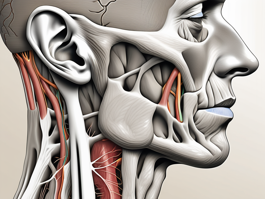
The greater auricular nerve is a critical component of the human nervous system, playing a significant role in sensory perception. It runs alongside the external jugular vein, which is another important anatomical feature. Understanding the anatomy and function of these structures is crucial for medical professionals and researchers alike, as it can shed light on various clinical conditions and potential treatment options. This article delves into the intricacies of the greater auricular nerve and its relationship with the external jugular vein.
Understanding the Greater Auricular Nerve
The greater auricular nerve, also known as the auricular branch of the cervical plexus, is a sensory nerve that supplies the skin over the side of the head, including the ear, parotid gland, and surrounding areas. It originates from the cervical plexus, specifically the second and third cervical nerves. Its primary function is to provide sensation to the aforementioned areas, allowing for proper perception and interpretation of stimuli.
Anatomy and Function of the Greater Auricular Nerve
The greater auricular nerve arises from the posterior border of the sternocleidomastoid muscle, extending upwards towards the ear. Along its course, it often travels alongside the external jugular vein. This nerve mainly carries sensory fibers from the skin of the external ear, as well as the skin over the parotid gland and the angle of the mandible. It provides vital input for temperature, touch, and pain sensation in these areas.
In addition to its sensory role, the greater auricular nerve also has an important function in maintaining the integrity of the cutaneous circulation in the head and neck regions. It achieves this by regulating blood flow and vascular tone, contributing to the overall homeostasis of these regions.
The intricate network of blood vessels in the head and neck region requires precise regulation to ensure proper blood supply and oxygenation. The greater auricular nerve, with its close proximity to the external jugular vein, plays a crucial role in this process. It helps to maintain optimal blood flow to the skin over the ear, parotid gland, and angle of the mandible, ensuring that these areas receive the necessary nutrients and oxygen.
The Greater Auricular Nerve’s Role in Sensory Perception
Sensory perception is a complex process that involves the transmission and interpretation of various stimuli. The greater auricular nerve is instrumental in this process, as it carries sensory information from the skin of the external ear, parotid gland, and adjacent areas to the brain.
When we touch our ear or feel the warmth of the sun on our face, it is the greater auricular nerve that transmits these sensations to the brain for interpretation. The nerve fibers within the greater auricular nerve are specialized to detect different types of stimuli, such as pressure, temperature, and pain. This allows us to have a comprehensive sensory experience in the areas it innervates.
Any disruptions or injuries to the greater auricular nerve can lead to sensory disturbances in its innervated areas. Patients may experience altered sensation, such as numbness, tingling, or even pain. It is essential to recognize and address these symptoms promptly to prevent further complications.
In conclusion, the greater auricular nerve is a vital component of the sensory system in the head and neck region. Its intricate anatomy and function allow for proper sensation and perception in the areas it supplies. Understanding the role of this nerve can help healthcare professionals diagnose and manage conditions that affect its function, ensuring optimal sensory perception for patients.
The External Jugular Vein: A Close Neighbor
The external jugular vein, as its name suggests, is a major superficial vein located in the neck. It is a tributary of the subclavian vein and facilitates the return of blood from the head and neck regions back to the heart. Its proximity to the greater auricular nerve is of particular interest due to their close relationship.
Anatomy and Function of the External Jugular Vein
The external jugular vein typically arises by the confluence of the posterior auricular vein and the retromandibular vein. From there, it descends obliquely across the sternocleidomastoid muscle before joining the subclavian vein.
The primary function of the external jugular vein is to drain deoxygenated blood from the scalp, face, and neck regions. It acts as an important conduit for the venous return from these areas, bringing the blood one step closer to reoxygenation.
The Relationship Between the External Jugular Vein and the Greater Auricular Nerve
The external jugular vein and the greater auricular nerve share a close anatomical relationship, often running alongside each other. This proximity is not only relevant from a gross anatomical standpoint but also has potential clinical implications.
Knowledge of the relationship between these structures is crucial in various medical procedures. For example, when performing venous access or surgically exploring the neck, it is vital to take into account the course and position of the external jugular vein to avoid potential nerve damage.
The Pathway of the Greater Auricular Nerve
The greater auricular nerve follows a distinct pathway during its course from the cervical plexus to its target areas. Understanding this pathway is pivotal in comprehending its function and clinical significance.
Origin and Termination of the Greater Auricular Nerve
The greater auricular nerve originates from the second and third cervical nerves, which are part of the cervical plexus. Specifically, it arises from the ventral rami of these nerves in the posterior region of the sternocleidomastoid muscle.
The nerve typically terminates by branching into several smaller nerves that supply the skin over the ear, parotid gland, and angle of the mandible. These sensory branches ensure widespread innervation of the aforementioned areas.
The Greater Auricular Nerve’s Journey Alongside the External Jugular Vein
During its course, the greater auricular nerve often travels in close proximity to the external jugular vein. This parallel course accentuates the interaction between these important structures, both anatomically and functionally.
The journey of the greater auricular nerve alongside the external jugular vein allows for potential cross-talk and feedback between them. This close association underscores the interconnected nature of our bodily systems and highlights the need for comprehensive medical assessment and treatment.
Clinical Significance of the Greater Auricular Nerve
The greater auricular nerve’s clinical significance extends beyond its role in sensory perception. It is essential to explore and understand its implications in various medical conditions and procedures to ensure optimal patient care.
Common Injuries and Conditions Affecting the Greater Auricular Nerve
Despite its protected course, the greater auricular nerve is not exempt from injuries or conditions that can affect its function. Trauma, infection, nerve compressions, or even systemic diseases can potentially lead to compromised sensation in its innervated areas.
Patients experiencing symptoms such as altered sensation, numbness, or pain in the ear, parotid gland, or adjacent regions should seek medical attention promptly. A healthcare professional can perform a comprehensive evaluation and recommend appropriate diagnostic tests and treatment options.
Diagnostic Procedures and Treatment Options
The diagnosis and treatment of conditions affecting the greater auricular nerve require a multidisciplinary approach. After an initial evaluation, healthcare professionals may recommend various diagnostic procedures, such as imaging studies or peripheral nerve testing, to assess the extent and nature of the nerve injury or pathology.
Treatment options depend on the underlying cause and severity of the condition. They may range from conservative management, such as physical therapy or pain management strategies, to surgical interventions aimed at decompression or repair of the nerve.
It is crucial to note that every patient’s case is unique, and treatment decisions should be made in consultation with a qualified healthcare professional. Seeking medical advice in a timely manner can lead to better outcomes and improved quality of life.
Future Research Directions
Despite significant advancements in our understanding of the greater auricular nerve and its relationship with the external jugular vein, several unanswered questions and potential research avenues remain. Exploring these areas can help expand our knowledge and pave the way for new treatment approaches.
Unanswered Questions About the Greater Auricular Nerve
There are still gaps in our understanding of the detailed anatomy, physiology, and functions of the greater auricular nerve. Further research is needed to elucidate the specific mechanisms by which it contributes to sensory perception and maintains cutaneous circulation.
Additionally, clarifying the impact of various injuries, diseases, and treatment modalities on the greater auricular nerve can provide valuable insights into the prevention and management of related conditions.
The Potential for New Treatment Approaches
Future research efforts may focus on developing innovative treatment approaches for conditions affecting the greater auricular nerve. Harnessing our growing understanding of nerve regeneration, tissue engineering, and neuroplasticity can potentially lead to the discovery of novel therapeutic strategies.
However, it is essential to highlight that any new treatments or approaches should be rigorously studied and validated through clinical trials before being implemented on a broad scale. This emphasizes the importance of ongoing research and collaboration within the medical community.
Conclusion
In conclusion, the greater auricular nerve and the external jugular vein exhibit a close anatomical relationship of clinical significance. Understanding the anatomy, function, and relationship between these structures can guide medical professionals in diagnosing and managing various conditions affecting these areas. Continued research into the greater auricular nerve and its potential therapeutic implications holds promise for future advancements in the field of neurology and vascular medicine. To ensure optimal care, individuals experiencing symptoms related to these structures should seek medical advice from qualified healthcare professionals.

Leave a Reply