The greater auricular nerve is a critical component of the human nervous system. Understanding its anatomy and function is essential for healthcare professionals and researchers alike. In this comprehensive guide, we will delve into the intricacies of greater auricular nerve palpation, its importance, techniques, potential challenges, and safety measures.
Understanding the Anatomy of the Greater Auricular Nerve
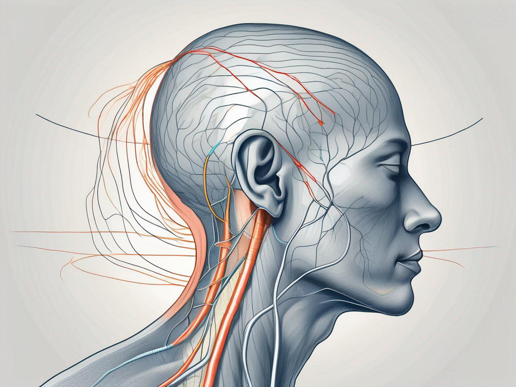
The greater auricular nerve is a fascinating structure that plays a crucial role in the sensory perception and innervation of the external ear, parotid gland, and adjacent skin regions. It originates from the cervical plexus and receives contributions from the second and third cervical nerves, forming an intricate network that allows for its diverse functions.
When examining the greater auricular nerve, healthcare providers must have a careful and detailed understanding of its pathway. By palpating the nerve, they can gain valuable insights into its health and potential underlying concerns, as well as assess its sensitivity and detect any abnormalities or signs of compromised sensory function.
Location and Function of the Greater Auricular Nerve
The greater auricular nerve runs a superficial course along the posterior border of the sternocleidomastoid muscle. It travels superiorly, providing sensory innervation to the skin overlying the parotid gland and ear, up to the level of the external acoustic meatus. This intricate pathway ensures that the nerve can effectively transmit sensory information from these regions to the central nervous system.
In addition to its sensory functions, the greater auricular nerve also plays a vital role in regulating blood flow and controlling vasomotor reflexes. This means that it not only provides us with the ability to perceive sensations but also contributes to the overall maintenance of our well-being.
The Role of the Greater Auricular Nerve in Sensory Perception
The sensory perception mediated by the greater auricular nerve is crucial for our everyday lives. It allows us to experience the world around us, from feeling the gentle touch of a breeze on our ears to sensing the warmth of the sun on our skin. Without the proper functioning of this nerve, our ability to perceive these sensations would be greatly compromised.
However, it is important to note that changes in sensory perception may be indicative of underlying medical conditions. For example, a decrease in sensitivity or the presence of abnormal sensations could be a sign of nerve damage or compression. Therefore, consulting with a healthcare provider is always recommended for an accurate diagnosis and appropriate treatment.
In conclusion, the greater auricular nerve is a remarkable structure that not only enables us to perceive sensory information but also contributes to the regulation of blood flow and vasomotor reflexes. Its intricate pathway and functions make it an essential component of our overall well-being. By understanding its anatomy and function, healthcare providers can effectively assess its health and address any concerns that may arise.
The Importance of Greater Auricular Nerve Palpation
Palpating the greater auricular nerve provides valuable information about its health and function. By incorporating this technique into clinical practice, healthcare professionals can identify potential nerve damage, evaluate pain sources, and implement targeted pain management strategies.
The greater auricular nerve, also known as the auricular branch of the vagus nerve, is a sensory nerve that supplies the skin over the external ear and the area just below it. It is responsible for transmitting sensations of touch, pain, and temperature from these regions to the brain. Palpating this nerve can help healthcare providers gain insights into its condition and functioning.
Identifying Nerve Damage through Palpation
Palpation of the greater auricular nerve can help detect possible nerve damage caused by trauma or other underlying conditions. By assessing the nerve’s texture, tension, and response to pressure, healthcare providers can gather essential diagnostic information.
During palpation, the healthcare professional gently applies pressure to the area where the nerve is located, usually just below the earlobe. They observe the patient’s response and note any abnormal sensations, such as tingling, numbness, or pain. These findings can indicate nerve compression, inflammation, or injury.
It is important to remember that nerve damage may manifest differently in different individuals, and a comprehensive evaluation by a medical professional should always be sought for an accurate diagnosis.
Palpation in Pain Management and Treatment
Effective pain management requires a multifaceted approach, and palpation of the greater auricular nerve can play a crucial role in this process. By identifying trigger points or areas of increased sensitivity, healthcare professionals can target these regions during treatment, whether through medications, physical therapy, or alternative therapies.
When palpating the greater auricular nerve, healthcare providers may discover tender points or areas of heightened sensitivity. These points can be indicators of nerve irritation or inflammation. By focusing treatment on these specific areas, healthcare professionals can provide targeted pain relief and improve overall patient outcomes.
It is essential to consult with a healthcare provider to develop an individualized treatment plan tailored to your specific needs and medical history. They can determine the most appropriate interventions based on the findings from greater auricular nerve palpation and other diagnostic assessments.
In conclusion, greater auricular nerve palpation is a valuable tool in healthcare practice. It allows for the identification of nerve damage and aids in the development of effective pain management strategies. By incorporating this technique into clinical assessments, healthcare professionals can provide more targeted and personalized care to their patients.
Techniques for Greater Auricular Nerve Palpation
To ensure accurate and effective palpation of the greater auricular nerve, healthcare providers must possess the appropriate knowledge, tools, and environment. Following a step-by-step guide, they can maximize the benefits of this technique.
The greater auricular nerve, also known as the auricular branch of the cervical plexus, plays a crucial role in innervating the skin over the external ear and the parotid gland. Palpating this nerve can provide valuable information about its health and function, aiding in the diagnosis and treatment of various conditions.
Preparing for Palpation: Necessary Tools and Environment
Before embarking on the palpation process, healthcare providers must gather the necessary tools, including gloves, sufficient lighting, and a tranquil environment that promotes patient comfort and relaxation.
Gloves are essential to maintain hygiene and prevent the transmission of pathogens. Adequate lighting is crucial for visualizing the anatomical landmarks and ensuring accurate palpation. Additionally, a tranquil environment helps create a sense of calmness, allowing the patient to feel more at ease during the procedure.
Creating a calm and supportive atmosphere not only enhances the effectiveness of the palpation but also helps alleviate any anxiety or apprehension the patient may experience. It is important for healthcare providers to establish open communication with the patient, addressing any concerns they may have and obtaining their informed consent before proceeding.
Step-by-Step Guide to Palpating the Greater Auricular Nerve
1. Begin by explaining the palpation process to the patient, addressing any concerns they may have. Obtain their informed consent before proceeding.
2. Put on gloves and ensure adequate lighting in the room. Position the patient comfortably, with their head turned slightly away from the side being examined.
3. Start by locating the sternocleidomastoid muscle and identifying its posterior border. The sternocleidomastoid muscle is a prominent muscle in the neck that can be easily palpated.
4. Gently palpate along the posterior border until you can feel a small, firm, and tubular structure, indicating the presence of the greater auricular nerve. The nerve is located superficially and can be felt as a distinct structure beneath the skin.
5. Assess the nerve’s texture, tension, and response to pressure. Note any asymmetry or areas of abnormal sensitivity. This information can provide valuable insights into the nerve’s health and function.
6. Communicate your findings to the patient and document the results for future reference. Effective communication ensures that the patient is well-informed about their condition and allows for appropriate treatment planning.
Remember, palpation should be performed by trained healthcare professionals who have a comprehensive understanding of the technique and its limitations. It is important to exercise caution and avoid applying excessive pressure during the palpation process to prevent any discomfort or injury to the patient.
By following these techniques for greater auricular nerve palpation, healthcare providers can gather valuable information about the nerve’s health and function, aiding in the diagnosis and treatment of various conditions. Palpation is a valuable tool in the hands of skilled professionals, contributing to comprehensive patient care and improved outcomes.
Potential Challenges and Solutions in Greater Auricular Nerve Palpation
While greater auricular nerve palpation can yield valuable insights, several challenges may arise during the process. Awareness of these challenges and having strategies to overcome them is key to successful palpation.
Common Difficulties in Palpating the Greater Auricular Nerve
Due to anatomical variations and individual differences, locating and palpating the greater auricular nerve may present challenges. Factors such as patient positioning, anatomical landmarks, and variations in nerve size can influence the palpation process.
Healthcare providers should adapt their approach accordingly and consider seeking guidance from experienced colleagues or specialists if challenges persist.
Tips and Tricks for Effective Palpation
1. Take the time to study and understand the anatomy of the greater auricular nerve before attempting palpation.
2. Seek additional training or guidance from experts to enhance your palpation skills.
3. Utilize gentle and gradual pressure during palpation to minimize patient discomfort.
4. Communicate with the patient throughout the process, ensuring their comfort and addressing any concerns they may have.
5. Maintain a comprehensive record of your findings and share them with the patient’s healthcare team for holistic care.
Safety Measures and Precautions in Greater Auricular Nerve Palpation
As with any medical procedure, ensuring patient safety is paramount during greater auricular nerve palpation. Healthcare providers must be aware of potential risks and adhere to necessary precautions.
Understanding the Risks of Incorrect Palpation
Improper palpation techniques can lead to patient discomfort, exacerbation of existing conditions, or potentially misleading diagnostic outcomes. Therefore, it is crucial for healthcare providers to receive adequate training and practice under supervision before performing greater auricular nerve palpation.
If you have concerns about your ability to perform this technique safely or professionally, consult with a healthcare provider who specializes in nerve assessment and palpation for expert guidance.
Ensuring Patient Comfort and Safety During Palpation
Patient comfort, trust, and safety should be prioritized throughout the palpation process. Ensure open communication with the patient, provide clear explanations, and address any discomfort they may experience during the procedure.
If a patient expresses apprehension or discomfort, consider alternatives or modifications to the technique and involve the patient in the decision-making process.
By following the techniques, understanding the challenges, and prioritizing patient safety, healthcare professionals can harness the power of greater auricular nerve palpation to improve patient care, pain management, and overall well-being.
Remember, always consult with a healthcare provider for an accurate diagnosis, individualized treatment plan, and further guidance regarding greater auricular nerve palpation.
Leave a Reply