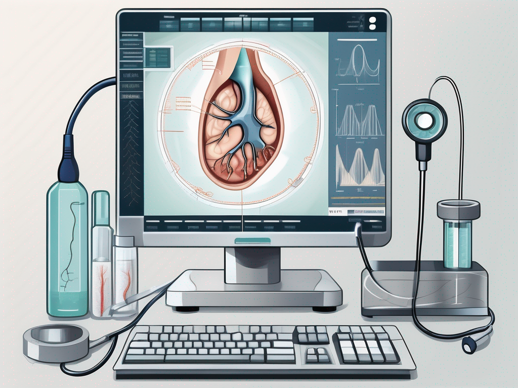
The Great Auricular Nerve, although not widely known, plays a crucial role in our health. In this comprehensive guide, we will explore the anatomy, function, and importance of the Great Auricular Nerve in medical diagnosis. We will also delve into the fascinating world of ultrasound technology and its role in assessing and monitoring the Great Auricular Nerve. By the end of this article, you will have a better understanding of the process, benefits, and limitations of Great Auricular Nerve Ultrasound.
Understanding the Great Auricular Nerve
Anatomy and Function of the Great Auricular Nerve
The Great Auricular Nerve is a branch of the cervical plexus, originating from the C2 and C3 nerve roots. It travels superficially along the side of the neck, providing sensory innervation to the external ear, parotid gland, mastoid region, and the skin over the angle of the mandible. This sensory pathway allows us to perceive touch, temperature, and pain in these areas.
The Great Auricular Nerve, with its intricate network of nerve fibers, plays a vital role in our daily lives. It is responsible for transmitting signals from the external environment to our brain, allowing us to react and respond accordingly. Without the Great Auricular Nerve, our ability to sense the world around us would be greatly diminished.
Furthermore, the Great Auricular Nerve works in conjunction with other nerves to help us maintain a healthy sensory perception. It communicates with neighboring nerves, sharing information and coordinating responses to various stimuli. This intricate network ensures that our sensory system functions optimally, allowing us to navigate our environment with ease.
Importance of the Great Auricular Nerve in Medical Diagnosis
Medical professionals rely on the Great Auricular Nerve as an indicator of potential underlying conditions. By assessing the function and integrity of this nerve, doctors can detect abnormalities or injuries that may be causing symptoms such as numbness, pain, or sensitivity in the head and neck areas.
Great Auricular Nerve Ultrasound stands as a valuable tool in diagnosing various medical conditions, such as nerve entrapment, trauma, inflammation, or tumors in the head and neck region. This non-invasive imaging technique allows healthcare providers to visualize the Great Auricular Nerve and assess its structure and function. By identifying any irregularities or abnormalities, medical professionals can develop targeted treatment plans to address the underlying cause of the patient’s symptoms.
Moreover, the Great Auricular Nerve serves as a crucial reference point for medical professionals during surgical procedures in the head and neck region. Surgeons must be mindful of the nerve’s location and function to avoid inadvertent damage during operations. By understanding the intricate anatomy and function of the Great Auricular Nerve, surgeons can perform procedures with precision and minimize the risk of complications.
In conclusion, the Great Auricular Nerve is a remarkable structure that plays a significant role in our sensory perception and overall well-being. Its intricate network of nerve fibers allows us to experience the world around us and helps medical professionals diagnose and treat various conditions. Understanding the importance of the Great Auricular Nerve highlights the complexity and interconnectedness of the human body, reminding us of the incredible wonders that lie within.
Introduction to Ultrasound Technology
Ultrasound technology is a revolutionary medical imaging technique that utilizes sound waves to create real-time images of the internal structures of the body. By using high-frequency sound waves, ultrasound provides valuable information about the body’s organs, tissues, and blood flow.
The process of ultrasound imaging involves the use of a transducer, which is a handheld device that emits sound waves. The transducer is placed against the skin and moved over the area of interest. As the sound waves travel through the body, they bounce off different tissues and organs, creating echoes. These echoes are then detected by the transducer and converted into images that can be seen on a screen.
One of the key advantages of ultrasound imaging is its non-invasive nature. Unlike other imaging techniques, such as X-rays or CT scans, ultrasound does not involve exposure to ionizing radiation. This makes it a safe method for both the patient and the healthcare provider.
Basics of Ultrasound Imaging
Ultrasound imaging is a versatile tool that can be used to examine various parts of the body, including the abdomen, pelvis, heart, blood vessels, and even the brain. It is commonly used to monitor the development of a fetus during pregnancy, diagnose conditions affecting the liver or gallbladder, and assess the function of the heart.
During an ultrasound examination, a gel is applied to the skin to ensure good contact between the transducer and the body. The transducer is then moved over the area of interest, capturing images in real-time. These images can provide valuable information about the size, shape, and texture of organs, as well as the presence of any abnormalities or diseases.
Ultrasound imaging is not only used for diagnostic purposes but also for guiding medical procedures. For example, it can be used to assist in the placement of needles during biopsies or to guide the drainage of fluid from a cyst or abscess.
Role of Ultrasound in Neurological Assessments
When it comes to the assessment of the Great Auricular Nerve, ultrasound plays a crucial role in visualizing and evaluating the nerve and its surrounding structures. The Great Auricular Nerve is a sensory nerve located in the neck and is responsible for providing sensation to the skin of the ear and the area behind the ear.
Using ultrasound, healthcare professionals can identify potential nerve compressions, tumors, inflammation, or other abnormalities that might be causing symptoms such as pain, numbness, or tingling in the ear or neck region. By visualizing the nerve in real-time, ultrasound allows for immediate and dynamic imaging, enabling healthcare professionals to assess the nerve’s function and integrity.
In addition to its diagnostic capabilities, ultrasound can also be used for therapeutic purposes. For example, ultrasound-guided nerve blocks can be performed to provide pain relief in patients with nerve-related conditions. By precisely targeting the affected nerve with the help of ultrasound imaging, healthcare providers can administer medication directly to the site of the problem, reducing pain and improving patient outcomes.
In conclusion, ultrasound technology has revolutionized the field of medical imaging, providing healthcare professionals with a safe, non-invasive, and real-time method of visualizing and assessing the internal structures of the body. Whether it is used for diagnostic purposes or guiding medical procedures, ultrasound plays a crucial role in improving patient care and outcomes.
The Process of Great Auricular Nerve Ultrasound
Preparing for the Ultrasound
Prior to the ultrasound procedure, it is important to follow any specific instructions given by your healthcare provider. These may include fasting for a certain period or refraining from applying any creams or lotions to the area being examined. It is also vital to inform the healthcare provider about any allergies or medical conditions you may have.
During the procedure, the patient is typically positioned comfortably with their head turned to the side. A gel is applied to the skin over the area of interest to help the transducer glide smoothly and improve the quality of the images.
Step-by-step Procedure of the Ultrasound
The healthcare provider will start by placing the transducer on the skin and gently moving it over the area being examined. They will then adjust various settings on the ultrasound machine to optimize the images captured.
As the transducer moves, an image will appear on the screen, showing the Great Auricular Nerve and surrounding structures in real-time. The healthcare provider will carefully assess the nerve’s size, shape, and any potential abnormalities.
The entire procedure usually takes around 15-30 minutes, depending on the level of detail required and the specific case being evaluated.
Interpreting Ultrasound Results
Reading and Understanding Ultrasound Images
Interpreting ultrasound images requires a skilled healthcare professional who is trained to recognize normal anatomy and identify any abnormalities. The images will typically display the Great Auricular Nerve, its course, and any surrounding structures.
During the evaluation, the healthcare provider will analyze the symmetry, size, and shape of the nerve, looking for any signs of compression, enlargement, inflammation, or masses.
Identifying Abnormalities and Potential Issues
If the imaging reveals any abnormalities or signs of nerve injury, the healthcare provider will discuss the findings with the patient. They will explain the potential causes and implications, outlining any necessary further investigations or treatments.
It is crucial to understand that ultrasound imaging alone may not provide a definitive diagnosis. Other tests or consultations with specialists may be required to confirm the findings and develop an appropriate treatment plan.
Benefits and Limitations of Great Auricular Nerve Ultrasound
Advantages of Using Ultrasound in Neurological Diagnosis
Great Auricular Nerve Ultrasound offers several advantages in the field of neurological diagnosis. It is a non-invasive, radiation-free imaging technique that provides real-time information about the nerve and its adjacent structures. This immediate feedback enables healthcare professionals to make timely and accurate diagnoses, leading to efficient management of patients’ conditions.
Furthermore, ultrasound is widely accessible, cost-effective, and well-tolerated by patients, making it a valuable tool in various healthcare settings.
Potential Challenges and How to Overcome Them
While ultrasound is a useful diagnostic tool, it does have its limitations. Factors such as operator experience, patient body habitus, and the location of the nerve can influence the quality and interpretability of the images.
To overcome these challenges, it is imperative to ensure that the healthcare provider performing the ultrasound is well-trained and experienced in neurosonography. Additionally, using advanced ultrasound machines and choosing the optimal scanning technique tailored to the individual patient can enhance the image quality and diagnostic accuracy.
If you believe you may benefit from Great Auricular Nerve Ultrasound, it is essential to consult with a medical professional who specializes in neurology or ultrasound. They will be able to assess your specific situation and guide you through the process, giving you the best possible care.
Conclusion
In this comprehensive guide, we have covered the basics of the Great Auricular Nerve, its anatomy, and its importance in medical diagnosis. We explored the world of ultrasound technology and its role in assessing the nerve’s function and integrity. Finally, we discussed the benefits and limitations of Great Auricular Nerve Ultrasound.
Remember, this article aims to provide information and raise awareness about Great Auricular Nerve Ultrasound. It is not a substitute for professional medical advice. If you are experiencing any symptoms or concerns related to your Great Auricular Nerve, please consult with a qualified healthcare professional who can provide appropriate guidance and care.
Leave a Reply