The great auricular nerve is an important structure in the human body, playing a crucial role in sensory function and providing valuable information for both clinical practitioners and researchers alike. Understanding the location and function of this nerve is paramount in various medical fields, such as surgery and pain management. In this comprehensive guide, we will delve into the intricate details of the great auricular nerve, exploring its anatomy, clinical importance, common disorders, and recent advances in research.
Understanding the Great Auricular Nerve
In order to grasp the significance of the great auricular nerve, it is essential to have a solid foundation of knowledge regarding its structure and function. Let us begin by exploring the anatomical aspects of this nerve.
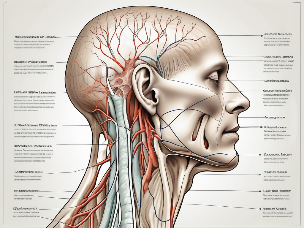
The great auricular nerve, also known as the auricular branch of the cervical plexus, plays a crucial role in the sensory innervation of the head and neck region. It originates from the spinal nerves C2 and C3, which are located in the upper part of the neck. From its origin, the great auricular nerve embarks on a complex journey, navigating its way through various anatomical structures.
As it ascends along the sternocleidomastoid muscle, the great auricular nerve gradually gains prominence, becoming more distinct and easily identifiable. This path allows the nerve to establish connections with other important structures in the neck, such as the lymph nodes and blood vessels.
Continuing its ascent, the great auricular nerve eventually reaches the superficial cervical fascia, a layer of connective tissue that envelops the neck. Here, it branches out and enters the parotid gland, one of the major salivary glands located in front of the ear. This branching pattern ensures that the nerve can distribute its sensory fibers to a wider area, maximizing its ability to transmit information.
Once inside the parotid gland, the great auricular nerve extends its reach, supplying sensory innervation to the skin overlying the ear and the surrounding area. This includes the region known as the parotid region, which encompasses the area in front of and below the ear, extending towards the angle of the mandible.
Now that we have explored the anatomical aspects of the great auricular nerve, let us delve into its function.
Anatomy of the Great Auricular Nerve
The great auricular nerve is a branch of the cervical plexus, stemming from the spinal nerves C2 and C3. It travels a complex path, ascending along the sternocleidomastoid muscle before reaching the superficial cervical fascia. From there, it enters the parotid gland and ultimately provides innervation to the skin overlying the ear and the surrounding area.
Function of the Great Auricular Nerve
The primary function of the great auricular nerve is sensory. It carries cutaneous sensation from the skin over the parotid region, external ear, and angle of the mandible. This information is then relayed to the brain, allowing for proper perception and interpretation of the external environment.
By transmitting sensory signals from the skin, the great auricular nerve enables us to perceive touch, temperature, and pain in the designated areas. It plays a crucial role in our ability to sense the external world and respond accordingly.
Moreover, the great auricular nerve is not only responsible for sensory innervation but also contributes to the autonomic nervous system. It participates in regulating blood flow, sweat production, and other involuntary functions in the region it supplies.
Understanding the great auricular nerve is essential for healthcare professionals, particularly those involved in head and neck surgery, as it allows for accurate diagnosis and treatment of various conditions affecting this nerve. By comprehending its anatomical course and function, medical practitioners can provide targeted interventions to alleviate pain, restore sensation, and improve overall patient outcomes.
Locating the Great Auricular Nerve
Accurately identifying the location of the great auricular nerve is of utmost importance, particularly in surgical procedures and pain management techniques. Let us explore the various ways to locate this nerve.
The great auricular nerve, also known as the auricular branch of the cervical plexus, is a sensory nerve that supplies the skin over the external ear and the angle of the mandible. It arises from the cervical plexus, which is a network of nerves formed by the anterior rami of the upper cervical spinal nerves.
Surface Landmarks for Identification
Locating the great auricular nerve can be guided by several surface landmarks. One common method involves identifying the sternoclavicular joint and measuring approximately 6 cm above it, tracing the course of the nerve along the sternocleidomastoid muscle.
The sternocleidomastoid muscle is a large muscle located in the anterior region of the neck. It originates from the sternum and clavicle and inserts into the mastoid process of the temporal bone. The great auricular nerve runs superficially along this muscle, making it a reliable landmark for identification.
Additionally, palpating the posterior border of the parotid gland serves as a reliable indicator for the entry point of the great auricular nerve into the gland. The parotid gland is the largest of the salivary glands and is located in front of the ear, extending into the neck. It produces saliva, which aids in the digestion of food.
When locating the great auricular nerve, it is essential to consider individual anatomical variations. The nerve’s course may vary slightly from person to person, and its position relative to the surface landmarks may differ. Therefore, a thorough understanding of the underlying anatomy is crucial for accurate identification.
Relation to Other Anatomical Structures
Understanding the relationship between the great auricular nerve and neighboring anatomical structures is vital in ensuring the success of surgical procedures and minimizing potential complications.
Notably, the nerve is at risk of damage during surgical interventions in the parotid region and certain reconstructive procedures, making careful dissection paramount. The parotid region is a complex anatomical area that houses important structures such as the facial nerve, which controls the muscles of facial expression, and the external carotid artery, which supplies blood to the face and scalp.
During parotid surgery, surgeons must exercise caution to avoid injuring the great auricular nerve, as damage to this nerve can result in sensory deficits and potential complications such as pain, numbness, and altered sensation in the external ear and the angle of the mandible.
Furthermore, reconstructive procedures involving the parotid region, such as facial nerve repair or tumor excision, require meticulous dissection to preserve the integrity of the great auricular nerve. Surgeons must navigate through the intricate network of nerves and blood vessels while ensuring the nerve’s protection and functionality.
In conclusion, accurate identification of the great auricular nerve is essential in various medical procedures. Surface landmarks, such as the sternoclavicular joint and the posterior border of the parotid gland, can guide the localization of this nerve. Understanding its relationship to neighboring anatomical structures is crucial to minimize complications and ensure successful outcomes. Surgeons must exercise caution and perform meticulous dissection when working in the parotid region to preserve the integrity of the great auricular nerve.
Clinical Importance of the Great Auricular Nerve
Recognizing the clinical significance of the great auricular nerve aids in optimizing patient care, particularly in surgical and pain management scenarios. Let us explore the various ways in which this nerve holds importance.
Role in Surgical Procedures
The great auricular nerve plays a crucial role in surgical procedures involving the parotid region and reconstructive interventions in the ear area. Its careful identification and preservation allow for improved patient outcomes and reduced postoperative complications.
Implications for Pain Management
Understanding the great auricular nerve’s role in pain perception offers valuable insights for pain management strategies. Nerve blocks and neuromodulation techniques can be employed to target this nerve specifically, providing relief for conditions such as trigeminal neuralgia and postoperative pain.
Common Disorders Involving the Great Auricular Nerve
Despite its importance, the great auricular nerve is susceptible to various disorders that can cause discomfort and impair its function. Recognizing the symptoms, undergoing proper diagnosis, and exploring appropriate treatment options are vital in managing these disorders effectively.
Symptoms and Diagnosis
Disorders involving the great auricular nerve may present with symptoms such as pain, numbness, tingling, or altered sensation over the area it innervates. Conducting a thorough clinical history, physical examination, and possibly imaging studies can aid in making an accurate diagnosis.
Treatment Options and Prognosis
Treatment options for disorders involving the great auricular nerve vary depending on the underlying cause and severity of symptoms. Conservative measures, such as pain medication and physical therapy, are often attempted initially. In more severe cases, surgical interventions or nerve blocks may be considered. The prognosis generally depends on early detection and appropriate management.
Recent Advances in Great Auricular Nerve Research
Ongoing research continues to shed light on the great auricular nerve, uncovering new insights into its functioning and potential clinical applications. Let us explore some recent advances that have the potential to revolutionize medical practice.
Innovations in Surgical Techniques
Surgical innovations, such as minimally invasive approaches and advanced imaging techniques, have greatly improved the accuracy and outcomes of procedures involving the great auricular nerve. These advancements aim to reduce procedural risks, optimize patient recovery, and enhance surgical precision.
New Insights into Nerve Function and Healing
Advancements in neurophysiology and regenerative medicine have granted researchers a deeper understanding of nerve function and healing mechanisms. This knowledge has the potential to open up new avenues for the treatment of nerve injuries, offering hope for improved patient outcomes and quality of life.
In conclusion, the great auricular nerve holds immense significance in numerous medical fields, ranging from surgery to pain management. Its accurate identification, understanding of anatomy, and recognition of clinical disorders are vital for healthcare professionals aiming to optimize patient care and outcomes. With ongoing research and advancements, the future holds promising prospects for further unlocking the mysteries of this remarkable nerve.
Leave a Reply