In the field of otolaryngology, parotideectomy refers to the surgical removal of the parotid gland, which is a major salivary gland located in the face. This surgical procedure is typically performed to treat various conditions such as tumors, infections, and stones within the gland. However, one potential complication that can occur after a parotideectomy is the development of a great auricular nerve neuroma.
Understanding Parotideectomy and Its Implications
The parotid gland plays a vital role in the production and secretion of saliva, contributing to the overall oral health and digestion of an individual. It is located near the ear, below the zygomatic arch, and extends towards the lower part of the face. During a parotideectomy, the gland is carefully dissected and removed, with the utmost precision and attention to surrounding structures.
The Role of the Parotid Gland
The parotid gland produces approximately 25% of the saliva in the oral cavity. It is responsible for lubricating the mouth, aiding in the digestion of food, and protecting against tooth decay. Additionally, the parotid gland also helps to regulate the pH levels in the mouth, thus preventing the growth of harmful bacteria.
Saliva, produced by the parotid gland, contains enzymes that initiate the breakdown of starches and fats in the mouth, facilitating the process of digestion. This gland also contributes to the overall taste sensation, as it secretes certain proteins that enhance the perception of flavors.
The Surgical Procedure of Parotideectomy
Parotideectomy is performed under general anesthesia and involves making an incision in front of or just below the ear. The surgeon then carefully removes the parotid gland while ensuring the preservation of vital structures such as the facial nerve. The duration of the surgery depends on the complexity of the case, and postoperative care is crucial to minimize complications and optimize recovery.
During the procedure, the surgeon meticulously dissects the parotid gland, taking care to avoid damage to the facial nerve. This nerve is responsible for controlling the muscles of facial expression and any injury to it can result in facial weakness or paralysis. To ensure the preservation of the facial nerve, the surgeon may use intraoperative nerve monitoring techniques, which provide real-time feedback on the nerve’s function.
Potential Post-Parotideectomy Complications
Although parotideectomy is a relatively safe procedure, there are potential complications that may arise. One such complication is the development of a great auricular nerve neuroma, a condition characterized by the abnormal growth or irritation of the great auricular nerve.
After a parotideectomy, some patients may experience sensory disturbances in the ear, face, and neck region. These disturbances may include numbness, tingling, or pain. These symptoms are typically transient, but in rare cases, they can persist and lead to the development of a neuroma.
In addition to sensory disturbances, patients may also experience temporary swelling and bruising around the surgical site. This is a normal part of the healing process and usually resolves within a few weeks. It is important for patients to follow their surgeon’s postoperative instructions, which may include the use of cold compresses and elevation of the head to reduce swelling.
Another potential complication of parotideectomy is the formation of a seroma, which is a collection of fluid that can accumulate in the surgical area. This can cause discomfort and delayed wound healing. To prevent the formation of a seroma, surgeons may place drains during the surgery to allow for the drainage of excess fluid.
It is important for patients to closely follow their surgeon’s instructions for postoperative care, including proper wound care and the use of prescribed medications. Regular follow-up appointments will also be scheduled to monitor the healing process and address any concerns or complications that may arise.
Great Auricular Nerve Neuroma: An Overview
The great auricular nerve is a sensory nerve that supplies the skin over the ear, cheek, and neck regions. A neuroma is an abnormal growth of nerve cells that can develop after a nerve injury or surgery. In the case of a parotideectomy, the manipulation or damage to the great auricular nerve during the procedure may lead to the formation of a neuroma.
Neuromas are fascinating entities that occur as a result of the body’s attempt to heal and regenerate damaged nerves. They consist of a disorganized collection of nerve fibers, scar tissue, and connective tissue. While they are noncancerous, they can cause a variety of symptoms and complications.
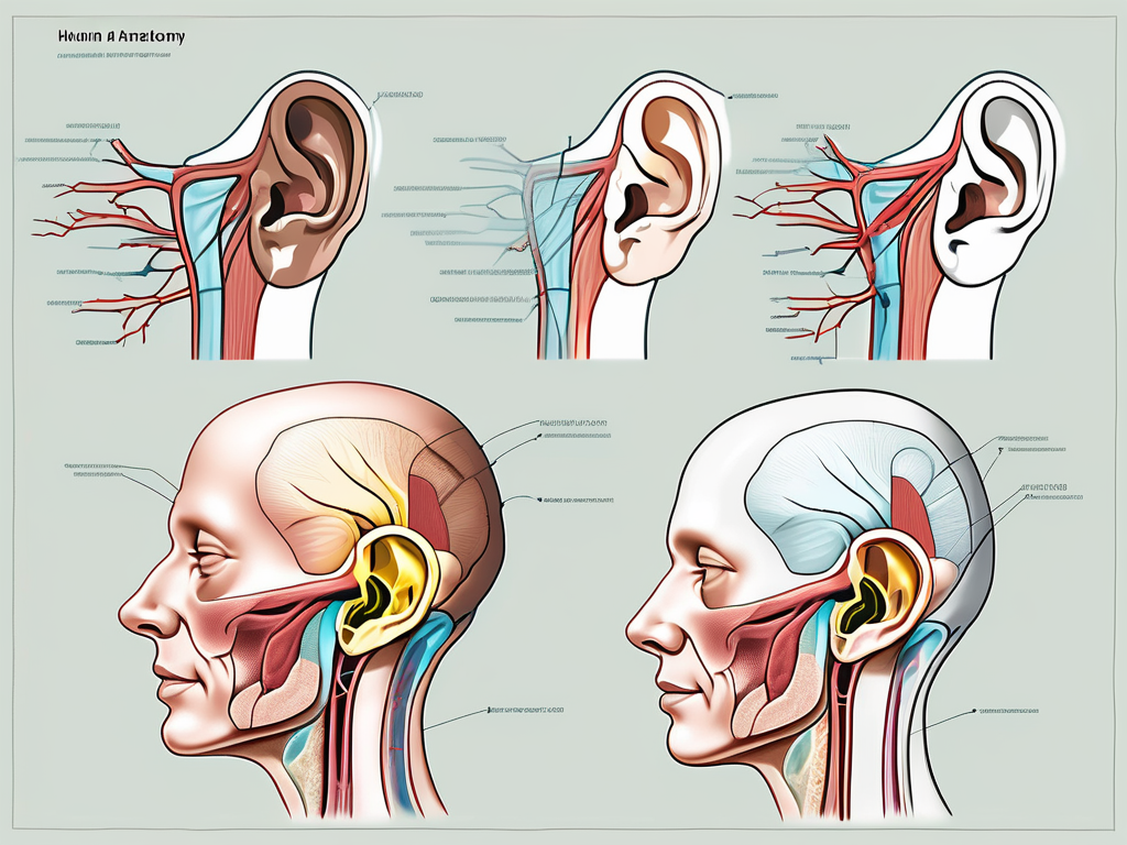
The great auricular nerve, originating from the cervical plexus, specifically from the spinal nerves C2 and C3, has a complex anatomical course. It travels along the posterior border of the sternocleidomastoid muscle, branching out to provide sensory innervation to the skin of the external ear, the parotid region, and parts of the neck. This intricate network of nerves allows us to perceive sensations and experience the world around us.
Anatomy of the Great Auricular Nerve
The great auricular nerve, like many other nerves in our body, is a marvel of nature’s design. It is composed of numerous individual nerve fibers, each with its own specific function and purpose. These fibers work together to transmit signals from the skin to the brain, allowing us to feel touch, temperature, and pain.
As the great auricular nerve courses its way through the body, it encounters various structures and tissues. It weaves its way through the intricate network of muscles, blood vessels, and connective tissue, ensuring that every inch of the ear, cheek, and neck regions is supplied with the necessary sensory input.
Defining Neuroma in Medical Terms
A neuroma, in simple terms, is an abnormal growth of nerve tissue. It occurs when the body’s natural healing process goes awry, resulting in the formation of a tangled mass of nerve fibers. This disorganized collection of nerve cells, scar tissue, and connective tissue can cause a range of symptoms and complications.
Neuromas are not limited to the great auricular nerve; they can develop in any nerve in the body. However, their occurrence in the great auricular nerve can be particularly troublesome due to the nerve’s extensive distribution and its role in providing sensory innervation to critical areas of the head and neck.
Symptoms and Diagnosis of Great Auricular Nerve Neuroma
The symptoms of a great auricular nerve neuroma can vary from person to person. While some individuals may experience pain or discomfort around the ear, cheek, or neck, others may notice altered sensation in the affected areas. In some cases, tenderness may be present upon palpation.
It is important to note that these symptoms may not solely result from a neuroma and can also be caused by other factors. Therefore, a thorough clinical examination is essential to determine the underlying cause of the symptoms. This examination includes a detailed history and physical examination, where the physician carefully evaluates the patient’s symptoms and performs various tests to assess nerve function.
In some cases, imaging studies such as MRI or ultrasound may be ordered to aid in confirming the diagnosis. These imaging modalities can provide detailed images of the affected area, allowing the physician to visualize any abnormalities or changes in the nerve structure. However, it is important to remember that the final diagnosis is typically made by a specialized physician based on a combination of clinical findings and imaging results.
The Connection Between Parotideectomy and Great Auricular Nerve Neuroma
Although relatively uncommon, the occurrence of a great auricular nerve neuroma has been reported following parotideectomy. This connection can be attributed to the inadvertent injury or manipulation of the great auricular nerve during the surgical procedure.
Surgical Risks Leading to Neuroma
During a parotideectomy, the surgeon must navigate through complex anatomical structures in close proximity to the parotid gland. Despite the utmost care and precision, there is a risk of accidental injury to the great auricular nerve. This unintended nerve injury can initiate the development of a neuroma.
The Incidence Rate of Neuroma Post-Parotideectomy
It is important to note that the occurrence of a great auricular nerve neuroma following a parotideectomy is considered a rare complication. The exact incidence rate is not well-documented and may vary depending on multiple factors including patient characteristics, surgical technique, and surgeon experience.
Treatment Options for Great Auricular Nerve Neuroma
Effective management of a great auricular nerve neuroma requires a multi-disciplinary approach involving various treatment modalities. The choice of treatment depends on factors such as the severity of symptoms, impact on daily activities, and the individual’s overall health and preferences.
Non-Surgical Interventions
In some cases, the symptoms associated with a great auricular nerve neuroma may be mild and may not significantly affect the individual’s quality of life. In such situations, conservative management options such as pain medications, physical therapy, and nerve blocks may be recommended. However, it is ultimately essential to consult with a healthcare professional to determine the most appropriate course of action.
Surgical Approaches to Neuroma Management
In cases where the symptoms are severe or conservative measures have been unsuccessful, surgical intervention may be considered. Surgical options for treating a great auricular nerve neuroma include excision of the neuroma, nerve grafting, or other nerve repair techniques. These procedures are generally conducted by specialists in peripheral nerve surgery to ensure optimal outcomes.
Rehabilitation and Recovery Process
Following surgical intervention, a comprehensive rehabilitation program is often recommended to aid in the recovery and restoration of function. This may include physical therapy, occupational therapy, and other complementary therapies tailored to the individual’s specific needs. Rehabilitation can play a crucial role in maximizing functional outcomes and improving overall quality of life.
In conclusion, while parotideectomy is a valuable surgical procedure for various conditions, the development of a great auricular nerve neuroma is a potential complication that should be considered. It is essential for individuals undergoing parotideectomy to be aware of this possibility and closely follow postoperative care instructions to minimize the risk of complications. If any symptoms suggestive of a neuroma arise, promptly consulting with a healthcare professional with expertise in otolaryngology can lead to early diagnosis and appropriate management strategies.
Leave a Reply