The human body is a complex system of interconnected nerves, each playing a vital role in our daily functions. One nerve that has generated considerable interest among researchers and medical professionals is the posterior auricular nerve. In this article, we will delve into the topic and explore whether the posterior auricular nerve is indeed a component of the facial nerve.
Understanding the Facial Nerve
Before we delve into the specifics of the posterior auricular nerve, it is crucial to have a solid understanding of the facial nerve itself. The facial nerve is one of the twelve cranial nerves that emerge directly from the brain. It is responsible for controlling the muscles of facial expression, as well as innervating certain glands and facilitating the sensation of taste on the anterior two-thirds of the tongue.
The facial nerve, also known as the seventh cranial nerve, is a complex and intricate structure that plays a vital role in our daily lives. It is responsible for allowing us to express our emotions through facial movements, such as smiling, frowning, and raising our eyebrows. Without the facial nerve, our faces would be devoid of expression, making it difficult for others to interpret our feelings and intentions.
Anatomy of the Facial Nerve
The facial nerve originates from the brainstem, specifically the pons, and travels through a narrow bony canal called the facial canal. Along its course, the facial nerve gives rise to several branches that supply different areas of the face. These branches include the temporal, zygomatic, buccal, marginal mandibular, and cervical branches.
The temporal branch of the facial nerve is responsible for innervating the muscles that raise the eyebrows and assist in closing the eyelids. The zygomatic branch controls the muscles involved in smiling, while the buccal branch controls the muscles of the cheeks and mouth. The marginal mandibular branch is responsible for the movements of the lower lip, and the cervical branch innervates the muscles of the neck.
It is fascinating to observe how the facial nerve branches out and supplies different regions of the face, each with its own unique function. This intricate network ensures that our facial expressions are precise and nuanced, allowing us to convey a wide range of emotions and communicate effectively with others.
Functions of the Facial Nerve
The facial nerve is primarily involved in controlling facial movements, allowing us to smile, frown, and express ourselves. Additionally, it plays a crucial role in tear and saliva production through its connections with the lacrimal and salivary glands. These glands are responsible for keeping our eyes moist and our mouths lubricated, ensuring optimal comfort and function.
Furthermore, the facial nerve enables us to taste the flavors we experience on the anterior two-thirds of our tongue. This sensory function is essential for our enjoyment of food and beverages, as well as our ability to discern different tastes and flavors. Without the facial nerve, our sense of taste would be significantly impaired, depriving us of one of life’s simple pleasures.
It is remarkable to think about the multifaceted role that the facial nerve plays in our daily lives. From allowing us to express our emotions to ensuring our eyes stay moist and our taste buds remain active, the facial nerve is truly a remarkable structure that deserves our appreciation and understanding.
The Posterior Auricular Nerve Explained
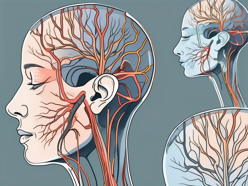
Now, let us shift our focus to the posterior auricular nerve. This nerve is a branch of the facial nerve, originating after the facial nerve exits the facial canal. The posterior auricular nerve innervates the muscles behind the ear, such as the occipitalis muscle and the auricular muscles.
The posterior auricular nerve, also known as the auricular branch, plays a crucial role in the intricate network of nerves that control the movements and sensations of the head and face. It is responsible for transmitting signals from the brain to the muscles located behind the ear, allowing for precise and coordinated movements.
Understanding the anatomy of the posterior auricular nerve is essential in comprehending its functions and significance. The nerve starts from the main trunk of the facial nerve and traverses behind the ear, where it supplies the aforementioned muscles. It then continues its course, innervating the scalp and the skin over the mastoid process.
Anatomy of the Posterior Auricular Nerve
The posterior auricular nerve arises from the facial nerve, which is one of the twelve cranial nerves. The facial nerve emerges from the brainstem and travels through a bony canal within the skull known as the facial canal. After exiting the facial canal, the facial nerve gives rise to several branches, including the posterior auricular nerve.
As the posterior auricular nerve makes its way behind the ear, it branches out to supply the occipitalis muscle and the auricular muscles. The occipitalis muscle, located at the back of the head, is responsible for retracting the scalp. The auricular muscles, on the other hand, control the movements of the external ear, allowing for subtle adjustments in its position and shape.
Continuing its journey, the posterior auricular nerve extends beyond the muscles it innervates and reaches the scalp. It provides sensory innervation to the scalp, allowing for the perception of touch, pressure, and temperature. Additionally, the nerve supplies the skin over the mastoid process, a prominent bony prominence located just behind the ear.
Functions of the Posterior Auricular Nerve
The primary function of the posterior auricular nerve is to control the movements of the muscles located behind the ear. It helps in movements related to the scalp and the auricles, contributing to facial expressions and the perception of sound.
When the brain sends signals through the posterior auricular nerve, the occipitalis muscle contracts, causing the scalp to retract. This movement is particularly noticeable when expressing surprise or raising the eyebrows. The auricular muscles, innervated by the posterior auricular nerve, allow for subtle adjustments in the position and shape of the external ear, aiding in the localization of sound sources.
In addition to its motor functions, the posterior auricular nerve also carries sensory information from the scalp and the skin over the mastoid process. This allows for the perception of various sensations, including touch, pressure, and temperature. The nerve endings present in these areas send signals back to the brain, providing valuable feedback about the external environment.
Overall, the posterior auricular nerve plays a vital role in the intricate network of nerves that control the movements and sensations of the head and face. Its branches supply important muscles behind the ear, contributing to facial expressions and the perception of sound. Additionally, the nerve provides sensory innervation to the scalp and the skin over the mastoid process, allowing for the perception of touch, pressure, and temperature.
The Connection Between the Facial Nerve and the Posterior Auricular Nerve
Understanding the connections and relationships between different nerves in the body is crucial to grasp the concept of whether the posterior auricular nerve is a component of the facial nerve.
The facial nerve, also known as cranial nerve VII, is one of the twelve cranial nerves that emerge directly from the brain. It is responsible for controlling the muscles of facial expression, as well as transmitting taste sensations from the anterior two-thirds of the tongue. The facial nerve emerges from the brainstem and passes through a small bony canal within the skull called the internal auditory meatus. From there, it branches out to innervate various muscles of the face.
The posterior auricular nerve, on the other hand, is a branch of the facial nerve that specifically innervates the muscles located behind the ear. It arises from the main trunk of the facial nerve and follows a distinct trajectory to reach its target muscles. This nerve provides motor innervation to the occipitalis muscle, which is responsible for moving the scalp backward.
Anatomical Proximity and Connections
The facial nerve and the posterior auricular nerve are closely positioned and share a common origin. They both arise from the facial nerve trunk, which is located deep within the parotid gland. The parotid gland is the largest of the salivary glands and is situated in front of the ear. The close proximity of these nerves allows for efficient communication and coordination between them.
As the facial nerve branches out, it gives rise to several other nerves, including the posterior auricular nerve. This branching pattern allows for the distribution of nerve fibers to different regions of the face and head. The posterior auricular nerve, being a branch of the facial nerve, shares a common origin with it but follows its own distinct pathway.
Shared Functions and Interactions
Although the facial nerve and the posterior auricular nerve have some overlapping functions, such as control of certain facial muscles, their primary functions and anatomical paths differ.
The facial nerve primarily governs facial expression, allowing us to smile, frown, raise our eyebrows, and make various other facial movements. It also plays a crucial role in transmitting taste sensations from the anterior two-thirds of the tongue to the brain. Damage to the facial nerve can result in facial paralysis or weakness, affecting the ability to make facial expressions and causing difficulties in speaking, eating, and drinking.
The posterior auricular nerve, on the other hand, has a more specific role in controlling the muscles behind the ear. It provides motor innervation to the occipitalis muscle, which is responsible for moving the scalp backward. This muscle is involved in various movements of the scalp, such as wrinkling the forehead and raising the eyebrows.
While the facial nerve and the posterior auricular nerve have their own distinct functions, they do interact and work together to some extent. For example, when we raise our eyebrows, both the facial nerve and the posterior auricular nerve are involved in coordinating the movement of the muscles responsible for this action.
In conclusion, the facial nerve and the posterior auricular nerve are closely related but have their own distinct functions and anatomical paths. Understanding their connections and interactions is essential for comprehending the complex network of nerves that control facial expression and movement.
Debates and Controversies in Neuroanatomy
The intricate nature of neuroanatomy often gives rise to debates and controversies. The question of whether the posterior auricular nerve is a component of the facial nerve has been the subject of discussion among experts.
Different Perspectives on Nerve Classification
Some experts argue that due to their anatomical proximity and shared origin, the posterior auricular nerve is indeed a component of the facial nerve. They highlight the close relationship between these nerves and how they work together to control facial expressions and related movements.
Implications for Neurological Disorders and Treatments
The ongoing debates surrounding the relationship between the posterior auricular nerve and the facial nerve have significant implications for the field of neurology. Understanding these connections may aid in the diagnosis and treatment of certain neurological disorders that affect facial muscle control and facial symmetry.
Conclusion: Is the Posterior Auricular Nerve a Component of the Facial Nerve?
In conclusion, while the posterior auricular nerve branches off from the facial nerve and shares some functions, it is not considered a component of the facial nerve itself. Its distinct anatomy, function, and innervation patterns differentiate it from the facial nerve. However, further research and advances in neuroanatomy may provide deeper insights into this complex relationship. If you have concerns or questions regarding any neurological matters, we strongly advise consulting with a qualified healthcare professional who can offer a thorough evaluation and appropriate guidance.
Leave a Reply