As someone interested in understanding the intricate workings of the human body, you may have come across the term “Greater Auricular Nerve”. In this comprehensive guide, we will delve into the anatomy, functions, clinical significance, treatment options, and prevention strategies related to this fascinating nerve. Before we begin, it’s important to note that while this article provides valuable information, it is not a substitute for professional medical advice. If you have any concerns regarding your health, it is always best to consult with a qualified healthcare professional.
Understanding the Greater Auricular Nerve
Anatomy of the Greater Auricular Nerve
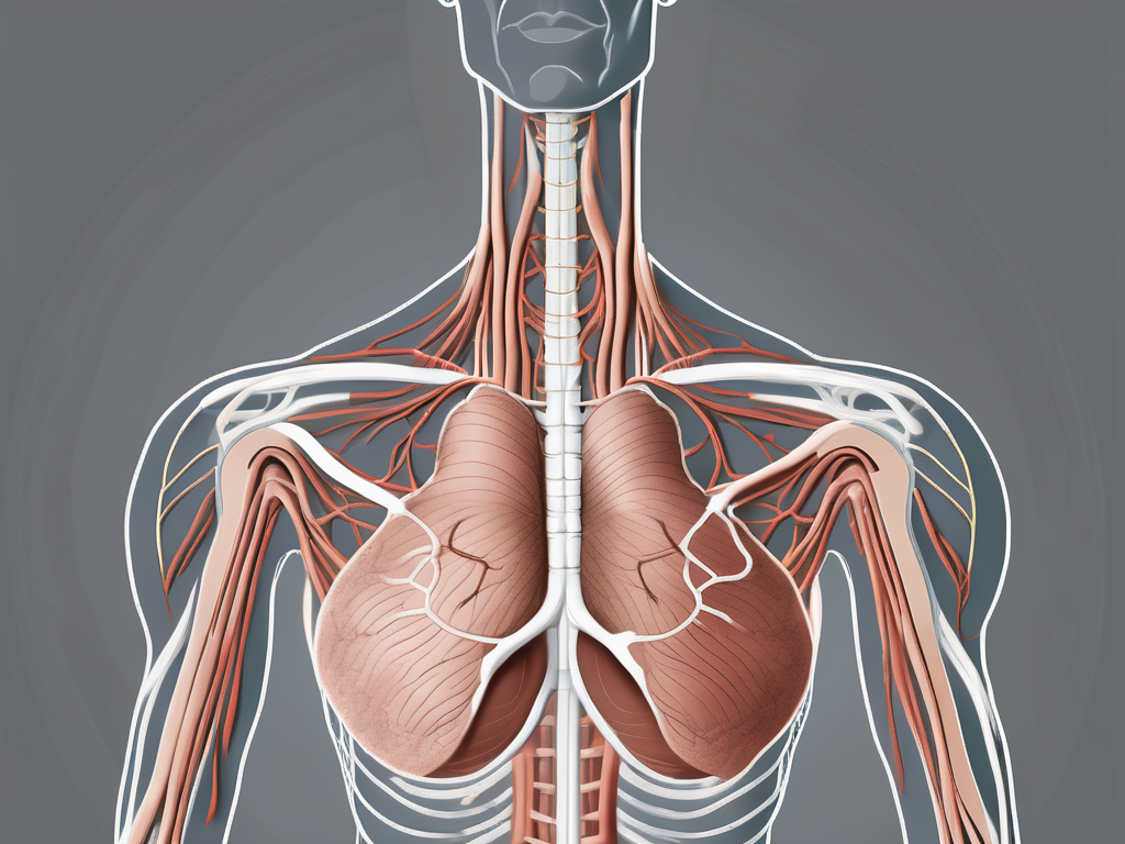
The Greater Auricular Nerve, also known as the auricular branch of the cervical plexus, is a sensory nerve that originates from the second and third cervical nerve roots (C2 and C3). It plays a crucial role in providing sensory feedback from the skin of the external ear to the brain, allowing us to perceive touch, pain, temperature, and other sensations in this region.
As the Greater Auricular Nerve travels along the posterior border of the sternocleidomastoid muscle, which is located in the neck near the ear, it forms a complex network of nerves in this region. It communicates with the lesser occipital nerve and the transverse cervical nerve, contributing to the intricate sensory pathways of the neck and ear.
The pathway of the Greater Auricular Nerve continues as it crosses the external jugular vein and ascends towards the ear. Along its course, it provides sensory innervation to the skin over the external ear, including the lobule, helix, and triangle of the auricle. This intricate network of nerve fibers ensures that the external ear is capable of perceiving various tactile sensations.
As the Greater Auricular Nerve nears the ear, it concludes its course by splitting into multiple branches that supply different areas of the ear. These branches further enhance the sensory capabilities of the external ear, allowing for a detailed perception of touch, pain, and temperature in specific regions.
Functions of the Greater Auricular Nerve
The Greater Auricular Nerve plays a crucial role in sensation, providing feedback from the skin of the external ear to the brain. This sensory information allows us to perceive touch, pain, temperature, and other sensations in this region. Understanding the functions of this nerve is essential in recognizing potential issues that may arise.
By understanding the anatomy and functions of the Greater Auricular Nerve, healthcare professionals can accurately diagnose and treat conditions that may affect this nerve. Common issues that may arise include nerve entrapment, trauma, inflammation, or compression. These conditions can lead to symptoms such as pain, numbness, tingling, or altered sensation in the external ear.
Furthermore, the Greater Auricular Nerve’s close proximity to other nerves in the neck and ear region highlights the importance of a comprehensive approach to diagnosis and treatment. Healthcare professionals must consider the interconnected nature of these nerves to ensure accurate assessment and management of any potential issues.
Research continues to uncover the intricate details of the Greater Auricular Nerve’s anatomy and functions, shedding light on its role in various medical conditions. By expanding our knowledge of this nerve, we can improve our understanding of the complex sensory pathways in the neck and ear, leading to more effective treatments and interventions.
The Pathway of the Greater Auricular Nerve
Origin and Course of the Greater Auricular Nerve
As mentioned earlier, the Greater Auricular Nerve arises from the C2 and C3 nerve roots of the cervical plexus. This complex network of nerves originates from the spinal cord, specifically the upper cervical vertebrae. From its origin, the Greater Auricular Nerve begins its remarkable journey.
After its formation, the nerve descends deep to the sternocleidomastoid muscle, a prominent muscle in the neck responsible for various movements of the head and neck. As the Greater Auricular Nerve travels beneath this muscle, it provides innervation to the skin on its posterior border, ensuring that this area remains sensitive to touch and other sensory stimuli.
The nerve then takes a superficial course, emerging from beneath the sternocleidomastoid muscle and passing diagonally across its surface. This superficial pathway allows the nerve to be more accessible and vulnerable to external factors. However, the body has developed protective mechanisms to safeguard the integrity of this vital nerve.
Continuing its journey, the Greater Auricular Nerve heads towards its ultimate destination – the ear. This remarkable organ responsible for hearing and maintaining balance is intricately connected to the nervous system, and the Greater Auricular Nerve plays a crucial role in ensuring its proper function.
Branches and Distribution of the Greater Auricular Nerve
Upon reaching the ear, the Greater Auricular Nerve divides into several branches, each with a specific mission to fulfill. These branches work harmoniously to supply the ear with sensory innervation, allowing us to fully experience the vibrant world of sound.
One of the branches covers the upper part of the ear, known as the helix. This branch delicately weaves its way through the intricate folds and contours of the helix, ensuring that every sensation is captured and transmitted to the brain for interpretation. The helix, with its unique shape and structure, plays a crucial role in capturing and funneling sound waves into the ear canal, where they can be transformed into meaningful auditory information.
Another branch of the Greater Auricular Nerve extends to the earlobe, providing sensation to this delicate area. The earlobe, often adorned with various accessories and cultural significance, is not only a visual focal point but also a sensory hotspot. The nerve endings in the earlobe allow us to appreciate the gentle touch of earrings or the warmth of a loved one’s hand resting on our ear.
Additionally, the Greater Auricular Nerve distributes branches to the area behind the ear, known as the mastoid region. This region, characterized by its bony prominence, serves as an attachment site for various muscles involved in head and neck movements. The nerve’s presence in this area ensures that any sensory information originating from the mastoid region is accurately transmitted to the brain, contributing to our overall perception of the surrounding environment.
Understanding the intricate distribution of the Greater Auricular Nerve is of utmost importance for medical professionals. This knowledge allows them to diagnose and treat potential issues associated with this region, ensuring that any disruptions to the nerve’s function are addressed promptly and effectively. By unraveling the pathway of the Greater Auricular Nerve, we gain a deeper appreciation for the complexity and interconnectedness of our nervous system.
Clinical Significance of the Greater Auricular Nerve
The Greater Auricular Nerve is a significant nerve that plays a crucial role in the sensory innervation of the ear and surrounding areas. It arises from the cervical plexus, specifically from the C2 and C3 spinal nerves. This nerve provides sensation to the skin over the parotid gland, the external ear, and the angle of the mandible.
While the Greater Auricular Nerve generally functions without issue, certain conditions may affect its proper functioning. One example is “neuralgia”, which refers to nerve pain. In some cases, individuals may experience sensitivity or discomfort in the distribution area of the Greater Auricular Nerve due to this condition. This can manifest as sharp, shooting pain or a constant dull ache. Neuralgia can be caused by various factors, including nerve compression, inflammation, or damage.
Other potential conditions that can affect the Greater Auricular Nerve include inflammation, infections, trauma, or entrapment of the nerve. Inflammation can occur as a result of an immune response or as a secondary effect of an underlying condition. Infections, such as cellulitis or abscesses, can lead to localized inflammation and subsequent nerve irritation. Trauma, such as direct injury or surgical procedures in the neck or ear region, can also cause damage to the Greater Auricular Nerve. Nerve entrapment, which occurs when the nerve is compressed or pinched, can result from factors like muscle spasms or anatomical abnormalities.
Diagnostic Techniques for Greater Auricular Nerve Disorders
When individuals present with symptoms that suggest a disorder of the Greater Auricular Nerve, medical professionals typically begin with a thorough examination. This may involve assessing the patient’s medical history, conducting a physical examination, and utilizing diagnostic tools such as imaging studies or nerve conduction tests.
During the medical history assessment, the healthcare professional will inquire about the onset, duration, and characteristics of the symptoms. They may also ask about any recent trauma, infections, or medical conditions that could be contributing to the nerve disorder. Additionally, a review of the patient’s medications and previous treatments may provide valuable insights.
The physical examination will focus on the affected area, including the ear, parotid gland, and mandible. The healthcare professional will carefully inspect the skin for any signs of inflammation, infection, or trauma. They may also perform sensory tests to evaluate the patient’s perception of touch, temperature, and pain in the distribution area of the Greater Auricular Nerve.
In some cases, imaging studies such as ultrasound, magnetic resonance imaging (MRI), or computed tomography (CT) scans may be ordered to visualize the nerve and surrounding structures. These imaging techniques can help identify any anatomical abnormalities, such as nerve compression or entrapment.
Nerve conduction tests, including nerve conduction velocity (NCV) and electromyography (EMG), may also be utilized to assess the function of the Greater Auricular Nerve. These tests involve the application of small electrical currents to the nerve and recording the resulting electrical activity. By measuring the speed and strength of the nerve impulses, healthcare professionals can evaluate the integrity and functionality of the nerve.
It is crucial to remember that only qualified healthcare professionals should perform these assessments to ensure accurate diagnosis and appropriate treatment. Depending on the underlying cause of the Greater Auricular Nerve disorder, treatment options may include medication for pain management, physical therapy, nerve blocks, or in severe cases, surgical intervention.
Treatment Options for Greater Auricular Nerve Disorders
Non-Surgical Treatments
In the majority of cases, conservative, non-surgical treatments are the first line of action for addressing Greater Auricular Nerve disorders. These non-invasive options may include medications to alleviate pain, anti-inflammatory treatments, physical therapy, or nerve blocks. The specific treatment plan will depend on the underlying condition and the recommendations provided by the healthcare professional overseeing the patient’s care.
Surgical Treatments
In some instances, surgical intervention may be necessary to address certain Greater Auricular Nerve disorders. Surgery might involve removing any obstacles that impede the nerve’s functionality or repairing damaged portions of the nerve. As with any surgical procedure, the decision for surgical intervention should be carefully considered and discussed with a healthcare professional.
Prevention and Management of Greater Auricular Nerve Disorders
Lifestyle Changes for Better Nerve Health
While it may not be possible to prevent all Greater Auricular Nerve disorders, certain lifestyle changes can promote overall nerve health. Maintaining a balanced diet, regular exercise, limiting exposure to stressors, and prioritizing good sleep hygiene are all factors that may indirectly contribute to nerve wellness.
Regular Check-ups and Early Detection
It is essential to prioritize regular check-ups and consultations with healthcare professionals to detect any potential issues with the Greater Auricular Nerve early on. Seeking medical advice for any concerning symptoms or changes in sensation around the ear can facilitate early intervention and improve outcomes.
Conclusion
Understanding the path of the Greater Auricular Nerve is crucial in recognizing and managing potential issues related to this important sensory nerve. While this comprehensive guide provides valuable insights, it is important to remember that only qualified healthcare professionals can provide personalized advice and treatment options. If you have any concerns regarding your health or the function of the Greater Auricular Nerve, it is advisable to seek professional medical assistance.
Leave a Reply