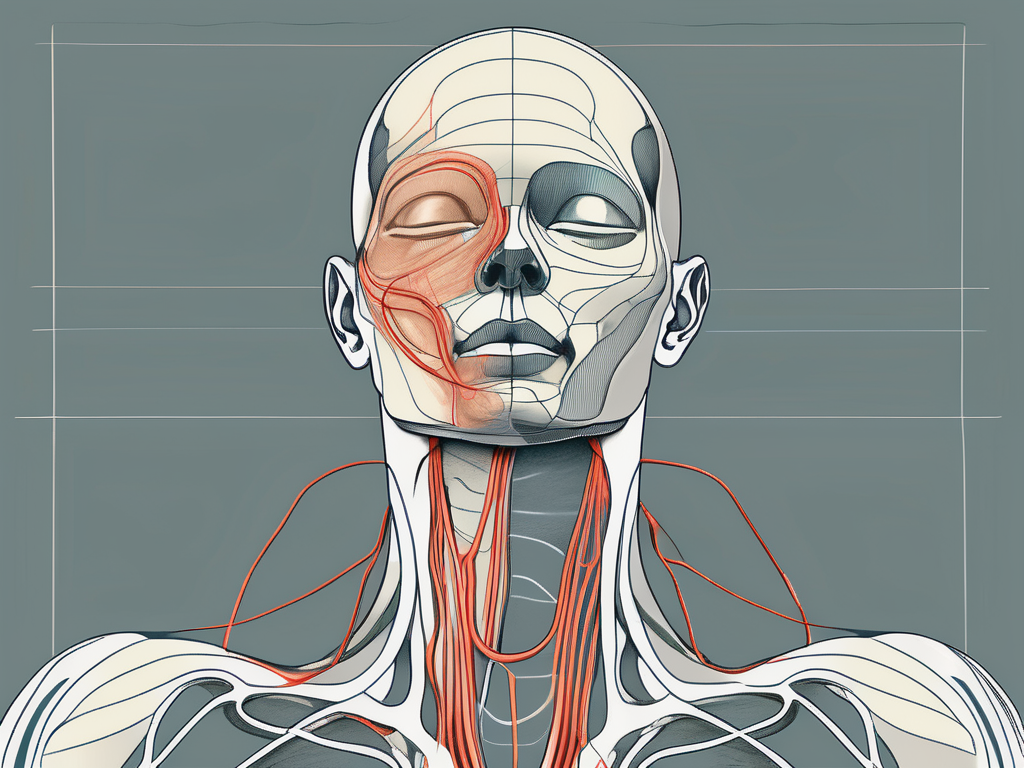
The suboccipital release technique is a commonly used therapy in the field of manual therapy and chiropractic care. It is often employed to alleviate tension and discomfort in the upper cervical region. However, some concerns have been raised regarding the potential for suboccipital release to pinch the greater auricular nerve, resulting in nerve damage and related complications. In this article, we will delve into the intricacies of the suboccipital release technique, explore the anatomy and function of the greater auricular nerve, discuss the potential risks and complications associated with suboccipital release, and provide insights into prevention and treatment options.
Understanding the Suboccipital Release Technique
The suboccipital release technique is a manual therapy that focuses on releasing tension and restoring mobility in the suboccipital region. The suboccipital region consists of a group of four muscles located at the base of the skull: the rectus capitis posterior major, rectus capitis posterior minor, obliquus capitis superior, and obliquus capitis inferior. These muscles play a crucial role in neck movement and stability.
When these suboccipital muscles become tight or restricted, they can cause a range of symptoms including headaches, neck pain, and limited range of motion. The suboccipital release technique aims to address these issues by targeting the specific muscles in this region.
The Anatomy of the Suboccipital Region
The suboccipital region is a highly complex and delicate area of the body. It serves as a bridge between the skull and the cervical spine, allowing for crucial movements such as head rotation and flexion. Additionally, this region houses important neurovascular structures, including the greater auricular nerve.
The rectus capitis posterior major muscle is located deep within the suboccipital region and is responsible for extending the head and neck. The rectus capitis posterior minor muscle, also deep within the region, assists in head rotation. The obliquus capitis superior muscle aids in head extension and rotation, while the obliquus capitis inferior muscle helps with head flexion.
Understanding the intricate anatomy of the suboccipital region is essential for a skilled practitioner to effectively perform the suboccipital release technique. By having a comprehensive knowledge of the muscles, nerves, and blood vessels in this area, the practitioner can ensure a safe and targeted approach to releasing tension and restoring mobility.
The Process of Suboccipital Release
During a suboccipital release, a skilled practitioner applies gentle pressure and manipulative techniques to the suboccipital muscles, aiming to release tension and restore normal function. The goal is to alleviate pain and improve range of motion in the neck and upper cervical region.
The practitioner may use their hands to apply specific techniques such as sustained pressure, stretching, or myofascial release to the suboccipital muscles. These techniques help to relax the muscles, increase blood flow, and promote healing in the area.
In addition to manual techniques, the practitioner may also incorporate other modalities such as heat therapy or ultrasound to further enhance the effects of the suboccipital release. These adjunct therapies can help to reduce inflammation, increase circulation, and promote relaxation in the suboccipital region.
It is important to note that the suboccipital release technique should only be performed by a trained and qualified practitioner. They will have the knowledge and skills to assess the individual’s condition, determine the appropriate techniques, and ensure the safety and effectiveness of the treatment.
Overall, the suboccipital release technique is a valuable manual therapy that can provide relief for individuals experiencing suboccipital muscle tension and related symptoms. By understanding the anatomy of the suboccipital region and following a systematic approach, practitioners can help restore mobility and improve overall well-being for their patients.
The Greater Auricular Nerve Explained
The greater auricular nerve is a branch of the cervical plexus, stemming from the second and third cervical nerves. It provides sensory innervation to the skin overlying the ear, the parotid gland, and the lateral portion of the scalp. Its intricate network of sensory fibers makes it susceptible to compression or damage in cases of trauma or excessive mechanical pressure.
Location and Function of the Greater Auricular Nerve
The greater auricular nerve travels superficially, running along the posterior border of the sternocleidomastoid muscle. It then ascends toward the ear, branching out to innervate various sensory areas. The main function of the greater auricular nerve is to provide sensory information from these regions to the brain, allowing for appropriate motor responses and perception of touch, warmth, and pain.
As the nerve courses along the sternocleidomastoid muscle, it sends branches to the skin overlying the ear, providing sensation to this delicate area. This sensory input allows us to feel the gentle touch of a loved one’s hand on our ear, the warmth of the sun’s rays, or the pain of an accidental bump.
In addition to the ear, the greater auricular nerve also supplies sensory innervation to the parotid gland, which is a major salivary gland located in front of the ear. This allows us to perceive any discomfort or pain in this area, which may indicate an underlying issue with the gland.
The lateral portion of the scalp is another region innervated by the greater auricular nerve. This includes the area above and behind the ear, extending towards the back of the head. The nerve fibers in this region transmit sensory information, enabling us to feel sensations such as the wind blowing through our hair or the touch of a hat on our scalp.
Common Issues and Disorders of the Greater Auricular Nerve
Injury or compression of the greater auricular nerve can lead to a variety of symptoms, including numbness, tingling, and pain in the affected areas. This can significantly impact an individual’s quality of life, as it may interfere with daily activities and overall well-being. Common disorders associated with the greater auricular nerve include entrapment neuropathy, injury due to trauma, and nerve compression caused by excessive pressure.
Entrapment neuropathy occurs when the nerve becomes trapped or compressed, leading to symptoms such as pain, numbness, and tingling. This can happen due to various factors, including prolonged pressure on the nerve, repetitive movements, or anatomical abnormalities.
Trauma, such as a direct blow to the ear or head, can also result in injury to the greater auricular nerve. This can cause immediate symptoms or develop over time, depending on the severity of the trauma. In some cases, surgical intervention may be required to repair the damaged nerve and restore function.
Excessive pressure on the nerve, often from activities such as wearing tight headbands or helmets, can lead to nerve compression. This can cause symptoms similar to entrapment neuropathy, including pain, numbness, and tingling. Avoiding activities that put undue pressure on the nerve and using protective equipment can help prevent this condition.
In conclusion, the greater auricular nerve plays a crucial role in providing sensory innervation to the ear, parotid gland, and lateral portion of the scalp. Understanding its location, function, and common issues can help individuals recognize and address any problems that may arise, ensuring optimal sensory perception and overall well-being.
The Connection Between Suboccipital Release and the Greater Auricular Nerve
While suboccipital release is primarily focused on addressing muscular tension and dysfunction, there is a potential for the technique to inadvertently affect the greater auricular nerve. The intricate proximity of the suboccipital region and the nerve path raises concerns regarding the possibility of nerve pinching or compression during the application of suboccipital release techniques.
How Suboccipital Release Can Affect the Greater Auricular Nerve
The precise mechanisms by which suboccipital release may impact the greater auricular nerve are not yet fully understood. However, it is plausible that direct mechanical pressure or indirect stretching of the nerve fibers during the release process could potentially compromise nerve function or induce symptoms related to nerve impingement or damage.
Potential Risks and Complications
It is important to note that reported cases of direct nerve damage resulting from suboccipital release are rare. However, it is crucial to be aware of the potential risks and complications associated with this technique. Symptoms such as worsened pain, tingling, numbness, or weakness after suboccipital release should be taken seriously and addressed promptly. Consulting with a healthcare professional, such as a primary care physician, neurologist, or chiropractor, is recommended to assess and manage any potential nerve-related issues.
Prevention and Treatment Options
Prevention of greater auricular nerve pinching during suboccipital release can be facilitated by employing appropriate techniques and a thorough understanding of the underlying anatomy. Skilled practitioners should ensure optimal patient positioning, apply gentle and controlled force, and constantly monitor patient feedback and response during the procedure to minimize the risk of nerve compression or damage.
Techniques to Avoid Nerve Pinching
Practitioners may employ various strategies to avoid nerve pinching during suboccipital release. These include using lighter pressure, modifying hand positions, or adjusting the direction and angle of the applied force. Additionally, the use of alternative techniques that do not directly apply pressure on the nerve region can be considered.
Therapeutic Approaches for Nerve Damage
In cases where nerve impingement or damage is suspected, therapeutic interventions may be employed to manage symptoms and promote nerve healing. These approaches can include physical therapy, medication management, and surgical intervention in severe or prolonged cases. Consulting with a healthcare professional is crucial to determine the most appropriate treatment plan based on individual circumstances.
The Role of Medical Professionals
When it comes to addressing concerns or issues related to the suboccipital region and the greater auricular nerve, the involvement of medical professionals is of utmost importance. Seeking consultation and guidance from healthcare providers with expertise in neurology, physical therapy, or chiropractic care is strongly advised.
When to Consult a Neurologist
If symptoms such as persistent pain, numbness, tingling, or weakness in the suboccipital or greater auricular nerve regions occur, it is prudent to consult with a neurologist. These specialists are trained to diagnose and treat nerve-related conditions, providing targeted interventions to address and manage the underlying causes of symptoms.
The Importance of a Skilled Physical Therapist
In cases where suboccipital release is recommended as part of a comprehensive treatment plan, the involvement of a skilled physical therapist can be invaluable. Physical therapists with expertise in manual therapy techniques, anatomy, and neurology can ensure optimal application of suboccipital release, minimizing the risk of complications and maximizing therapeutic benefits.
Ultimately, suboccipital release techniques can be effective in addressing tension and dysfunction in the suboccipital region. However, the potential for nerve pinching or compression, particularly of the greater auricular nerve, must be carefully considered. It is essential to work closely with qualified healthcare professionals and understand individual risk factors and treatment options. By prioritizing patient safety and employing appropriate techniques, the benefits of suboccipital release can be maximized, while minimizing the potential risks to the greater auricular nerve.

Leave a Reply