-
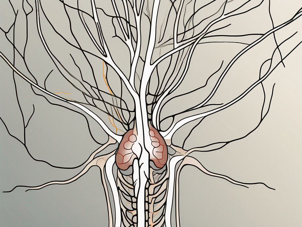
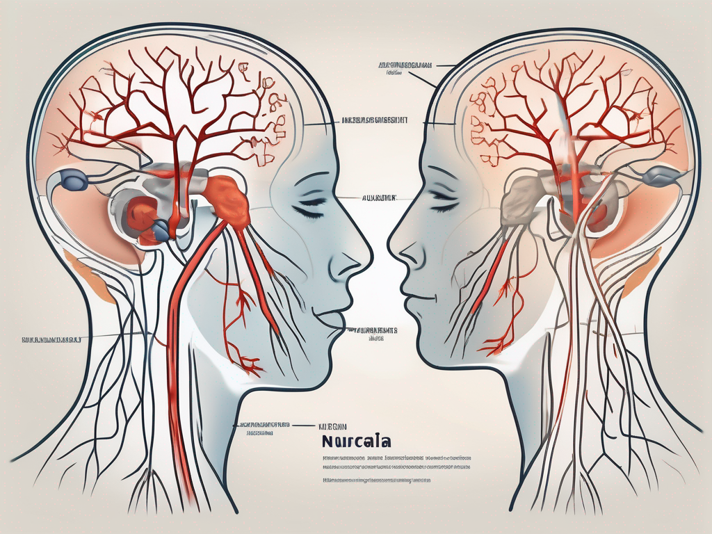
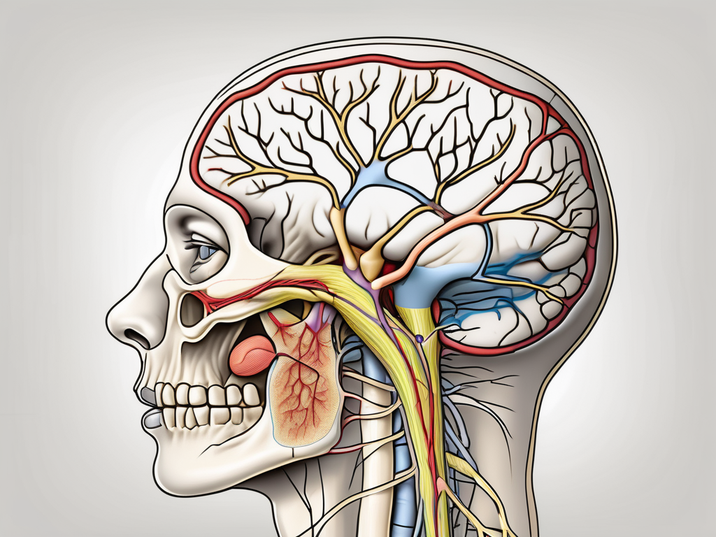
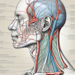
Which Cervical Nerve Does the Great Auricular Nerve Branch From?
Discover the intricate anatomy of the great auricular nerve as this article delves into the specific cervical nerve from which it branches.
-
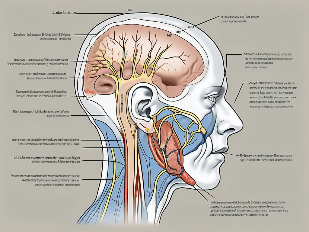
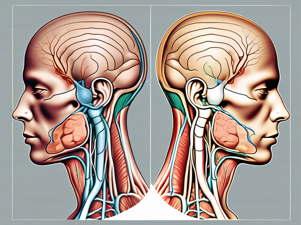
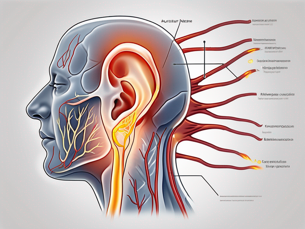
Understanding the Pre-Auricular Nerve: Functions, Disorders, and Treatments
Explore the intricate world of the pre-auricular nerve in this comprehensive article, delving into its vital functions, common disorders, and effective treatment options.
-
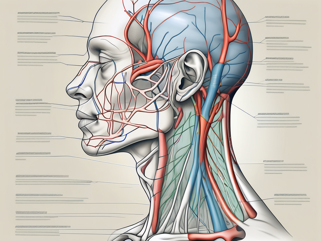
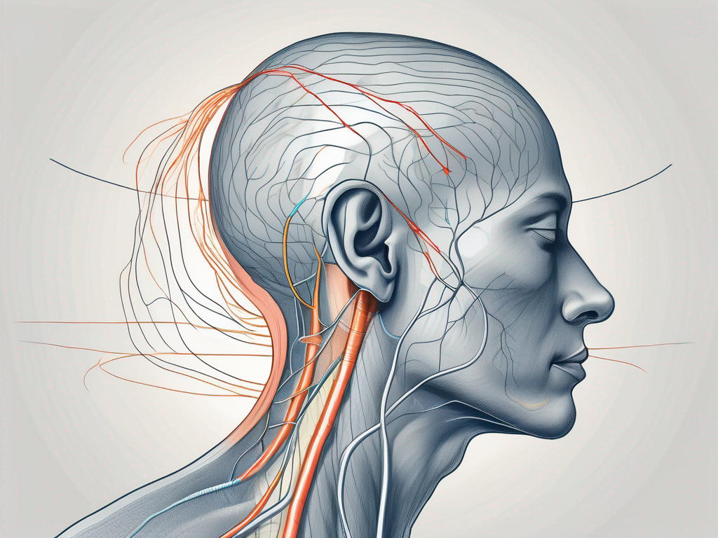
The Relationship Between the External Jugular Vein and Great Auricular Nerve
Explore the intricate connection between the external jugular vein and great auricular nerve in this fascinating article.
-
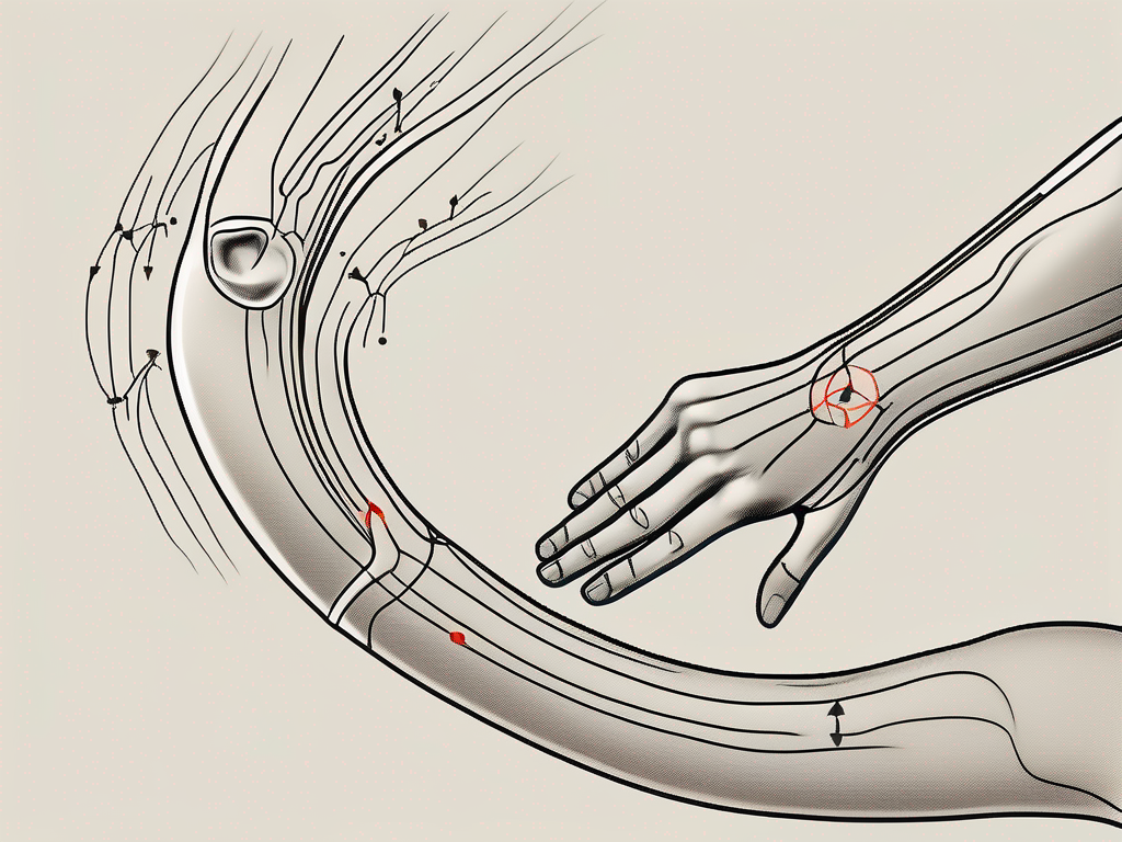
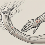
Understanding Pinched Great Auricular Nerve: Causes, Symptoms, and Treatment
Explore the causes, symptoms, and treatment options for a pinched great auricular nerve in this comprehensive article.
-
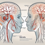
Understanding Greater Auricular Nerve Neuralgia: Causes, Symptoms, and Treatment
Discover the ins and outs of Greater Auricular Nerve Neuralgia in this comprehensive article.
-
Understanding the Great Auricular Nerve and Transverse Cervical Nerve: A Comprehensive Guide
Delve into the intricate world of the Great Auricular Nerve and Transverse Cervical Nerve with our comprehensive guide.
-
A Comprehensive Guide to Greater Auricular Nerve Palpation
Learn how to effectively palpate the greater auricular nerve with our comprehensive guide.
-
The Benefits of PNS and Auricular Nerve Stimulation: A Comprehensive Guide
Unlock the potential of PNS and Auricular Nerve Stimulation with this comprehensive guide.
-
The Origin of the Greater Auricular Nerve: Unveiling its Roots
Uncover the fascinating history and origins of the Greater Auricular Nerve in this enlightening article.
-
Can You Burn the Greater Auricular Nerve?
Discover the truth about the potential risks and consequences of burning the Greater Auricular Nerve.
Recent Posts
- Which Cervical Nerve Does the Great Auricular Nerve Branch From?
- Understanding the Pre-Auricular Nerve: Functions, Disorders, and Treatments
- The Relationship Between the External Jugular Vein and Great Auricular Nerve
- Understanding Pinched Great Auricular Nerve: Causes, Symptoms, and Treatment
- Understanding Greater Auricular Nerve Neuralgia: Causes, Symptoms, and Treatment
Tags
There’s no content to show here yet.