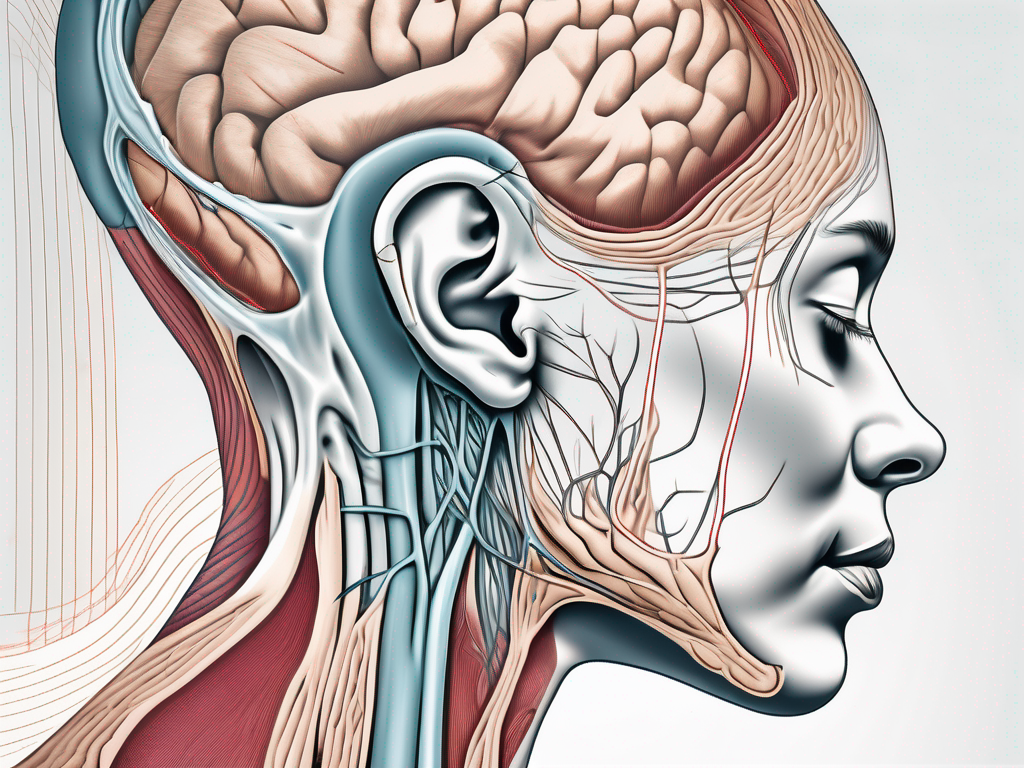
The Great Auricular Nerve is a crucial nerve located in the neck region. Understanding its anatomy and function is essential for various medical procedures, including anesthesia and surgical interventions. In this comprehensive guide, we will explore the different aspects of the Great Auricular Nerve, from its origin and pathway to techniques for locating it. We will also discuss common mistakes in identifying the nerve and safety measures to avoid nerve damage during procedures. Let’s dive in!
Understanding the Anatomy of the Great Auricular Nerve
The Great Auricular Nerve, also known as the posterior auricular nerve, originates from the cervical plexus. It arises from the second and third cervical nerves and travels upward, crossing over the sternocleidomastoid muscle. Its pathway is remarkable, as it courses behind the external ear, supplying sensation to the skin over the ear and the parotid gland.
The nerve consists of sensory fibers that transmit information about pain, temperature, and touch from the respective areas it innervates. Understanding the anatomy of the Great Auricular Nerve is crucial for medical professionals involved in procedures that may be affected by its distribution.
The Origin and Pathway of the Great Auricular Nerve
The Great Auricular Nerve originates from the cervical plexus, specifically from the second and third cervical nerves. It courses superiorly, traveling behind the sternocleidomastoid muscle. From there, it ascends towards the external ear, supplying sensory innervation to the skin overlying the ear and the parotid gland.
As the nerve travels behind the external ear, it sends branches to the skin, providing sensation to the area. These branches are responsible for the perception of pain, temperature, and touch in this region. The pathway of the Great Auricular Nerve is intricate, ensuring that the sensory information from the ear and parotid gland is transmitted accurately to the central nervous system.
The Function and Importance of the Great Auricular Nerve
The function of the Great Auricular Nerve is mainly sensory. It provides innervation to the skin over the ear, allowing for the perception of pain, temperature, and touch in this area. Additionally, it supplies sensation to the parotid gland, which is an important salivary gland located just in front of the ear.
The parotid gland plays a crucial role in the production of saliva, which aids in the digestion of food. The sensory fibers of the Great Auricular Nerve ensure that any changes in the parotid gland, such as inflammation or injury, are detected and transmitted to the brain for appropriate responses.
In medical procedures such as anesthesia and surgery, the knowledge of the Great Auricular Nerve’s anatomy and function is crucial. It helps medical professionals avoid potential complications associated with this nerve. By understanding the precise pathway and distribution of the Great Auricular Nerve, healthcare providers can minimize the risk of injury or damage during procedures that involve the ear or parotid gland.
Furthermore, the Great Auricular Nerve is often used as a landmark in surgical procedures. Its position and course provide a reliable reference point for surgeons, aiding in the identification and preservation of nearby structures. This knowledge is particularly important in surgeries involving the head and neck region, where precision and accuracy are paramount.
Techniques for Locating the Great Auricular Nerve
Locating the Great Auricular Nerve accurately is essential to prevent complications during procedures involving the neck and ear regions. Here are two common techniques used to locate this nerve:
Palpation Techniques
Palpation is a commonly used technique to locate the Great Auricular Nerve. By applying gentle pressure on specific anatomical landmarks, such as the sternocleidomastoid muscle and the external ear, medical professionals can identify the nerve’s position relative to these structures. This technique requires a thorough understanding of the anatomy and a skilled touch to accurately locate the nerve.
During palpation, the medical professional carefully feels for the nerve’s presence, noting any variations in its pathway. The sternocleidomastoid muscle, located on the side of the neck, serves as a useful landmark for identifying the nerve’s position. By palpating along the muscle, the medical professional can trace the nerve’s path as it branches out towards the ear.
Additionally, the external ear provides another reference point for locating the Great Auricular Nerve. By gently palpating the area around the ear, medical professionals can feel for the nerve’s presence and determine its proximity to the surface. This information is crucial for ensuring accurate nerve blocks or other procedures involving the nerve.
However, it is important to note that variations in nerve pathway may exist, so palpation alone may not always provide an accurate location. In cases where the nerve’s pathway differs from the norm, additional techniques may be necessary to ensure precise identification.
Ultrasound Guided Identification
Ultrasound technology has revolutionized medical procedures by providing real-time imaging of anatomical structures, including nerves. Using ultrasound guidance, medical professionals can accurately identify the Great Auricular Nerve. This technique offers a non-invasive and highly accurate method for locating the nerve, enhancing patient safety and procedural success rates.
During an ultrasound-guided identification, a small handheld device called a transducer is used to emit high-frequency sound waves into the body. These sound waves bounce back as echoes when they encounter different tissues, creating a real-time image on a monitor. By visualizing the nerve’s trajectory and avoiding any anatomical variations, medical professionals can precisely locate the Great Auricular Nerve.
Ultrasound-guided identification is especially useful in cases where palpation alone is not sufficient. It allows medical professionals to visualize the nerve’s position in relation to surrounding structures, such as blood vessels and muscles, ensuring accurate needle placement during nerve blocks or other procedures.
Furthermore, ultrasound technology provides the ability to assess the nerve’s size, shape, and any potential abnormalities. This information can aid in diagnosing conditions affecting the Great Auricular Nerve and determining the most appropriate treatment plan.
In conclusion, both palpation techniques and ultrasound-guided identification play crucial roles in locating the Great Auricular Nerve. While palpation relies on a skilled touch and knowledge of anatomical landmarks, ultrasound technology offers real-time imaging and precise visualization. By combining these techniques, medical professionals can ensure accurate identification of the nerve, enhancing patient safety and procedural success rates.
Common Mistakes in Identifying the Great Auricular Nerve
Identifying the Great Auricular Nerve can be challenging, and certain mistakes are commonly made. Being aware of these pitfalls is crucial for medical professionals to ensure successful procedures. Here are two common mistakes to avoid:
Misinterpretation of Anatomical Landmarks
One common mistake is misinterpreting anatomical landmarks. While palpation relies on identifying specific structures, variations in anatomy can lead to inaccuracies. For example, the position of the mastoid process, which is often used as a reference point, can vary among individuals. This variation can result in improper nerve localization, potentially leading to ineffective anesthesia or accidental nerve injury. Therefore, it is important for medical professionals to thoroughly understand the anatomical landmarks and their potential variations in order to accurately identify the Great Auricular Nerve.
Moreover, the surrounding structures, such as the sternocleidomastoid muscle and the external jugular vein, can sometimes obscure the Great Auricular Nerve. This can further complicate the identification process, as these structures may need to be carefully maneuvered or temporarily retracted to gain clear access to the nerve. Therefore, medical professionals should exercise caution and take their time when assessing the anatomical landmarks to avoid misinterpretation and potential complications.
Overlooking Variations in Nerve Pathway
The Great Auricular Nerve’s pathway can sometimes deviate from the textbook presentation. While the nerve is typically expected to travel along a predictable path, variations can occur. These variations may include changes in the nerve’s course, branching patterns, or even its proximity to other structures. Overlooking these variations can lead to unsuccessful nerve localization during procedures.
For instance, the Great Auricular Nerve may occasionally take a more superficial or deeper course than expected. This can make it challenging to identify and anesthetize the nerve accurately. Additionally, the nerve may exhibit branching patterns that differ from the standard anatomy, further complicating the identification process. Therefore, medical professionals must stay vigilant and adaptable, constantly reassessing the nerve’s pathway and adjusting their approach accordingly.
Furthermore, it is important to consider the potential impact of adjacent structures on the nerve’s pathway. For example, the presence of scar tissue, tumors, or inflammation in the surrounding area can alter the nerve’s course or make it more difficult to locate. By carefully evaluating the patient’s medical history, conducting thorough physical examinations, and utilizing imaging techniques when necessary, medical professionals can better anticipate and address these variations in the Great Auricular Nerve’s pathway.
Safety Measures in Handling the Great Auricular Nerve
Ensuring patient safety is paramount when handling the Great Auricular Nerve. Here are two important safety measures to consider:
Avoiding Nerve Damage During Procedures
During procedures involving the Great Auricular Nerve, it is essential to take precautions to avoid nerve damage. Careful dissection and manipulation of surrounding tissues, precise instrument handling, and adherence to established protocols can help minimize the risk of nerve injury. Always consult with a specialist and follow best practices to ensure patient safety.
Post-Procedure Care and Monitoring
After performing procedures involving the Great Auricular Nerve, it is crucial to provide adequate post-procedure care and monitoring. This includes proper wound care, monitoring for signs of infection or nerve injury, and addressing any patient concerns. If any complications arise, promptly consult with a healthcare professional to ensure appropriate management.
The Role of the Great Auricular Nerve in Medical Procedures
The Great Auricular Nerve plays a crucial role in various medical procedures, particularly those involving the neck and ear regions. Here are two areas where its involvement is significant:
Anesthesia and the Great Auricular Nerve
During anesthesia administration, the Great Auricular Nerve’s innervation should be considered to ensure adequate pain control for patients. By identifying and avoiding this nerve, anesthetists can tailor their techniques to provide effective anesthesia while minimizing the risk of unintended nerve blocks or complications. Always consult with an anesthesiologist to determine the best approach.
Surgical Procedures Involving the Great Auricular Nerve
In surgical procedures, such as plastic surgery or ear surgeries, the Great Auricular Nerve’s anatomical knowledge is crucial. Surgeons must take care to preserve the nerve’s integrity while performing their respective interventions. Accurate identification and proper handling of the nerve ensure functional outcomes and patient satisfaction.
In conclusion, understanding the Great Auricular Nerve’s anatomy, locating techniques, and potential pitfalls is vital for medical professionals involved in procedures related to the neck and ear regions. Always consult with a healthcare professional for accurate guidance and advice specific to individual patient needs. By prioritizing patient safety and staying up-to-date with advancements in nerve localization, medical professionals can carry out successful procedures and provide optimal care for their patients.

Leave a Reply