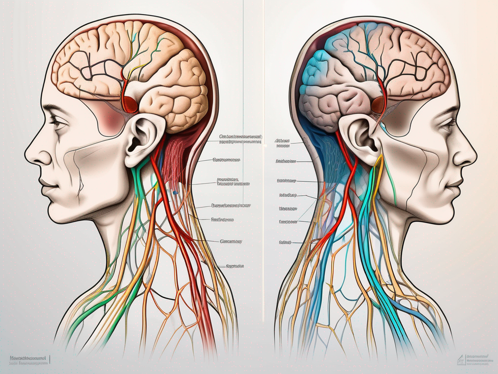
The auriculotemporal nerve and great auricular nerve are both important components of the human nervous system, playing crucial roles in sensory and motor functions. By understanding their anatomy, function, and clinical significance, we can gain valuable insights into the complexities of these nerves and appreciate their role in our overall well-being.
Anatomy of the Auriculotemporal Nerve
The auriculotemporal nerve is a significant nerve in the head and neck region. It plays a crucial role in the sensory innervation of various areas, including the face, temple, and external ear. Let’s delve deeper into the intricate details of this fascinating nerve.
Origin and Course of the Auriculotemporal Nerve
The auriculotemporal nerve originates from the trigeminal ganglion, which is a sensory ganglion located within the cranial cavity. Specifically, it arises from the mandibular division of the fifth cranial nerve, also known as the trigeminal nerve.
After its origin, the auriculotemporal nerve courses through the infratemporal fossa, which is a space located deep within the skull. It then emerges anteriorly, near the temporomandibular joint, which is the joint responsible for jaw movement.
From its emergence, the nerve branches out, spreading its sensory fibers to various regions of the face. These branches play a crucial role in transmitting sensory information from the skin to the brain.
Branches and Distribution of the Auriculotemporal Nerve
Once the auriculotemporal nerve emerges from its origin, it gives rise to several branches, each with its own unique distribution and function.
One of the main branches of the auriculotemporal nerve innervates the skin over the temple, providing sensory information related to touch, temperature, and pain in this region. This branch is responsible for the sensation you feel when you touch your temple or when the temperature changes in that area.
Another branch of the auriculotemporal nerve extends towards the auricle, which is the external part of the ear. This branch supplies sensory fibers to the skin of the auricle, allowing you to perceive touch, pressure, and pain in this region.
In addition to the temple and auricle, the auriculotemporal nerve also sends branches to adjacent regions, further expanding its sensory distribution. These branches ensure that the skin in these areas is adequately innervated, allowing for proper sensory perception and protection against potential harm.
In conclusion, the auriculotemporal nerve is a vital component of the trigeminal nerve system. Its origin, course, and distribution are intricately designed to provide sensory innervation to the face, temple, and external ear. Understanding the anatomy of this nerve helps us appreciate the complexity of the human body and its remarkable ability to sense and perceive the world around us.
Function of the Auriculotemporal Nerve
The auriculotemporal nerve serves both sensory and motor functions within the head and neck region.
The auriculotemporal nerve, a branch of the mandibular division of the trigeminal nerve, is a vital component of the sensory innervation of the face. It emerges from the infratemporal fossa and courses upward, passing through the parotid gland before branching out to supply various regions of the head and neck.
Sensory Functions
The sensory fibers of the auriculotemporal nerve play a crucial role in perceiving touch and temperature sensations from the skin of the temple, auricle, and adjacent regions. These sensory signals are essential for our ability to feel and interpret the world around us.
When you touch a hot surface, the sensory fibers of the auriculotemporal nerve are responsible for relaying the information to your brain, triggering a reflexive withdrawal response to protect you from potential harm. Similarly, when a gentle breeze brushes against your temple, it is the auriculotemporal nerve that allows you to experience the sensation of touch.
Furthermore, the sensory fibers of the auriculotemporal nerve also play a role in pain perception. In response to injury or inflammation in the temple and auricle regions, these fibers transmit pain signals to the brain, alerting you to potential harm or underlying health issues.
Motor Functions
While primarily a sensory nerve, the auriculotemporal nerve also contributes to motor functions. It carries parasympathetic fibers that facilitate salivation, specifically from the parotid gland.
The parotid gland, located just in front of the ear, is one of the major salivary glands in the human body. It produces saliva, which is crucial for the initial breakdown of food and the facilitation of swallowing. The parasympathetic fibers carried by the auriculotemporal nerve stimulate the parotid gland to produce saliva, ensuring proper oral health and aiding in the digestive process.
In addition to its motor function related to salivation, the auriculotemporal nerve also contributes to the regulation of blood flow in the head and face. It helps to dilate blood vessels in the temple region, allowing for increased blood flow when necessary, such as during physical exertion or exposure to cold temperatures.
Anatomy of the Great Auricular Nerve
The great auricular nerve is a crucial component of the cervical plexus, originating from the anterior rami of the second and third cervical nerves. This nerve plays a vital role in providing sensory innervation to specific areas of the head and neck.
One of the primary regions innervated by the great auricular nerve is the skin over the parotid gland and the auricle. This means that any touch, temperature, or pain sensations experienced in these areas are transmitted to the brain through the great auricular nerve.
Origin and Course of the Great Auricular Nerve
Deep within the neck, the great auricular nerve originates from the cervical plexus. It emerges anteriorly and superiorly, crossing over the sternocleidomastoid muscle, which is a prominent muscle responsible for various movements of the head and neck.
Once it crosses over the sternocleidomastoid muscle, the great auricular nerve begins its ascent towards the parotid gland. During this journey, it courses laterally along the face, following a distinct path that ensures its proper distribution of sensory fibers to the designated areas.
The intricate course of the great auricular nerve allows it to navigate through the complex anatomy of the neck and face, ensuring that it reaches its intended destination with precision.
Branches and Distribution of the Great Auricular Nerve
As the great auricular nerve travels towards the auricle and parotid gland, it gives off branches that supply sensory fibers to the skin over these regions. These branches play a crucial role in transmitting touch, temperature, and pain sensations to the brain, allowing us to perceive and respond to stimuli in these areas.
These sensory fibers are responsible for relaying valuable information to the brain, enabling us to distinguish between various tactile sensations and respond accordingly. Without the great auricular nerve and its branches, our ability to perceive touch, temperature, and pain in the auricle and parotid gland regions would be compromised.
Understanding the anatomy and function of the great auricular nerve provides valuable insights into the intricate network of nerves within the head and neck. It highlights the importance of this nerve in our sensory perception and serves as a reminder of the remarkable complexity of the human body.
Function of the Great Auricular Nerve
The great auricular nerve is primarily involved in sensory functions within the head and neck region.
Sensory Functions
The sensory fibers of the great auricular nerve provide crucial input for perceiving touch, temperature, and pain sensations from the skin over the parotid gland and auricle. They contribute to our overall sensory experience and are vital for our awareness of the external environment.
Motor Functions
Unlike the auriculotemporal nerve, the great auricular nerve does not have a significant role in motor functions. It primarily carries sensory information and does not contribute to motor control of any specific muscles or glands.
Clinical Significance of the Auriculotemporal and Great Auricular Nerves
The auriculotemporal and great auricular nerves can be involved in various clinical disorders and symptoms. It is essential to understand their clinical significance to recognize and address potential issues.
Common Disorders and Symptoms
Disorders that affect the auriculotemporal and great auricular nerves can lead to symptoms such as neuropathic pain, altered sensation, and hypersensitivity. Conditions like trigeminal neuralgia or neuralgia-inducing cavitational osteonecrosis (NICO) may impact the function of these nerves, resulting in significant discomfort and impaired quality of life.
Diagnostic Procedures
To evaluate the function of the auriculotemporal and great auricular nerves, diagnostic tests such as nerve conduction studies and electromyography may be conducted. These tests help assess nerve conduction velocity and identify any abnormalities or damage present.
Treatment Options and Prognosis
The treatment of disorders affecting the auriculotemporal and great auricular nerves depends on the underlying cause and severity of symptoms. Treatment options may include medications to manage pain and inflammation, physical therapy, or in severe cases, surgical interventions. It is essential to consult with a healthcare professional for an accurate diagnosis and appropriate treatment plan tailored to individual needs.
In conclusion, understanding the role of the auriculotemporal nerve and great auricular nerve is crucial in comprehending the complexities of the human nervous system. These nerves play essential roles in sensory and motor functions within the head and neck region. Recognizing their anatomy, function, and clinical significance allows us to appreciate their importance and address any potential issues that may arise. If you are experiencing any symptoms related to these nerves, it is advised to seek guidance from a healthcare professional to receive a proper diagnosis and appropriate treatment.

Leave a Reply