The auriculotemporal nerve and greater auricular nerve are two important branches of the trigeminal nerve that play distinct roles in sensory innervation of the head and neck. Understanding the differences between these nerves is crucial for healthcare professionals working in areas such as neurology, otolaryngology, and dentistry. In this article, we will explore the anatomy, functions, and clinical significance of the auriculotemporal nerve and greater auricular nerve, as well as discuss the similarities and differences between them.
Anatomy of the Auriculotemporal Nerve
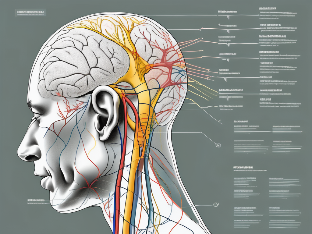
The auriculotemporal nerve is an important branch of the mandibular division of the trigeminal nerve (CN V3). It plays a crucial role in providing sensory innervation to various regions of the head, including the external ear, the skin of the temple, and the scalp posterior to the ear. Understanding the origin, pathway, and functions of the auriculotemporal nerve is essential for healthcare professionals in diagnosing and managing conditions related to this nerve.
Origin and Pathway of the Auriculotemporal Nerve
The auriculotemporal nerve arises from the main trigeminal ganglion located within the middle cranial fossa. This ganglion is a collection of nerve cell bodies that receive sensory information from the face and transmit it to the brain. From the trigeminal ganglion, the auriculotemporal nerve travels through a bony canal called the foramen ovale, which is located in the skull. This canal provides a protected pathway for the nerve to exit the skull and reach its target areas.
Once the auriculotemporal nerve exits the skull, it passes superiorly in front of the external auditory meatus, which is the opening of the ear canal. This path allows the nerve to provide sensory innervation to the external ear. As it continues its course, the nerve then divides into anterior and posterior branches. These branches further extend their reach, providing sensory innervation to the skin of the temple and the scalp posterior to the ear.
Functions of the Auriculotemporal Nerve
The auriculotemporal nerve carries general sensory fibers responsible for pain, temperature, and touch sensation. It plays a vital role in transmitting these sensations from the external ear, temple, and scalp posterior to the ear to the brain. This sensory information allows individuals to perceive and respond to various stimuli in their environment.
In addition to its sensory functions, the auriculotemporal nerve also carries parasympathetic fibers. These fibers provide secretomotor innervation to the parotid gland, which is one of the major salivary glands located in front of the ear. The parotid gland produces saliva, which aids in the digestion of food and maintaining oral health. The parasympathetic fibers of the auriculotemporal nerve stimulate the secretion of saliva, contributing to proper salivary gland function.
Clinical Significance of the Auriculotemporal Nerve
The auriculotemporal nerve plays a significant role in various clinical scenarios. Its involvement is often seen in temporomandibular joint (TMJ) disorders, which are conditions affecting the joint that connects the jawbone to the skull. Dysfunction of the TMJ can cause referred pain to the temple region, where the auriculotemporal nerve provides sensory innervation. This referred pain can be debilitating and may require medical intervention for relief.
Furthermore, the auriculotemporal nerve can be affected in conditions such as trigeminal neuralgia or cluster headaches. Trigeminal neuralgia is a condition characterized by severe facial pain, often triggered by simple activities such as eating or speaking. The involvement of the auriculotemporal nerve in this condition can lead to excruciating pain in the areas it innervates, including the temple and the scalp posterior to the ear. Cluster headaches, on the other hand, are severe headaches that occur in clusters or patterns. The involvement of the auriculotemporal nerve in cluster headaches can contribute to the intense pain experienced by individuals during these episodes.
Consultation with a healthcare professional experienced in the management of temporomandibular joint disorders, trigeminal neuralgia, or cluster headaches is essential for proper diagnosis and treatment. Understanding the anatomy and functions of the auriculotemporal nerve can aid healthcare professionals in providing appropriate care and relief to individuals experiencing pain or dysfunction related to this nerve.
Anatomy of the Greater Auricular Nerve
The greater auricular nerve is a branch of the cervical plexus, specifically originating from the second and third cervical nerves (C2 and C3). It begins its journey by traveling upward along the sternocleidomastoid muscle, a large muscle located in the neck. As it ascends, it crosses over the internal jugular vein, a major blood vessel in the neck, before branching into multiple smaller nerve fibers.
These smaller nerve fibers have specific destinations, providing sensory innervation to various areas. One important destination is the skin overlying the external ear, where the greater auricular nerve plays a crucial role in transmitting touch, pressure, and temperature sensations. Additionally, it also provides sensory innervation to the parotid gland, a salivary gland located in front of the ear, as well as the angle of the mandible, which is the junction of the lower jawbone.
Functions of the Greater Auricular Nerve
The primary function of the greater auricular nerve is to carry general sensory fibers responsible for transmitting touch, pressure, and temperature sensations from the innervated areas. It does not have any motor function, meaning it does not control any muscles or movement.
Specifically, the greater auricular nerve provides sensory innervation to the skin overlying the earlobe, cheeks, and the region just below the ear. This allows us to perceive and respond to various tactile stimuli in these areas, such as feeling the texture of an earring on the earlobe or sensing the warmth of sunlight on the cheek.
Clinical Significance of the Greater Auricular Nerve
The greater auricular nerve is of significant clinical importance, particularly in procedures involving the head and neck region. Surgeons and healthcare professionals often encounter this nerve during surgeries, such as earlobe reconstructions or other reconstructive procedures.
However, as with any nerve, injuries to the greater auricular nerve can occur, leading to potential complications. Damage to the nerve can result in altered sensation or numbness in the areas it innervates, including the skin overlying the earlobe, cheeks, and the region just below the ear. Patients who experience persistent or worsening symptoms, such as tingling, loss of sensation, or pain in these areas, should seek medical attention for further assessment and management.
It is important for healthcare professionals to be aware of the anatomical course and function of the greater auricular nerve to minimize the risk of injury during surgical procedures. Additionally, understanding the clinical significance of this nerve allows for appropriate management and treatment of any potential complications that may arise.
Comparing the Auriculotemporal and Greater Auricular Nerves
Similarities Between the Two Nerves
Both the auriculotemporal nerve and greater auricular nerve are branches of the trigeminal nerve with similar functions, providing sensory innervation to specific regions of the head and neck. They are both involved in transmitting general sensory information related to touch, temperature, and pain sensation. Additionally, they both have clinical significance and can be affected in various pathological conditions resulting in pain or altered sensation.
Differences in Function and Structure
While the auriculotemporal nerve carries parasympathetic fibers that provide secretomotor innervation to the parotid gland, the greater auricular nerve does not have any motor function. Additionally, the pathways and origins of these two nerves differ. The auriculotemporal nerve arises from the trigeminal nerve, while the greater auricular nerve originates from the cervical plexus. These differences in structure give rise to the variation in the regions they innervate and their clinical implications.
Implications for Medical Practice
Diagnostic Considerations
Understanding the anatomical distribution and functional differences between the auriculotemporal nerve and greater auricular nerve is vital in clinical settings. Healthcare professionals should consider the involvement of these nerves in cases of head and neck pain or altered sensation. Accurate diagnosis requires a detailed medical history, physical examination, and, if necessary, additional diagnostic tests such as imaging or nerve conduction studies. It is crucial to consult with a healthcare professional experienced in neurological disorders or oro-facial pain for appropriate assessment and management.
Treatment Approaches for Nerve-Related Conditions
Management of nerve-related conditions involving the auriculotemporal nerve and greater auricular nerve may vary depending on the underlying cause. Treatments can range from conservative measures such as oral medications for pain management to more invasive interventions like nerve blocks or surgical procedures. It is essential to seek guidance from a healthcare professional who specializes in nerve-related disorders to determine the most appropriate treatment options.
Future Research Directions
Unanswered Questions in Nerve Anatomy
Although we have made significant progress in understanding the anatomy and function of the auriculotemporal nerve and greater auricular nerve, several questions remain unanswered. Further research is needed to explore the precise mechanisms of pain transmission along these nerves and their interactions with other sensory pathways. Additionally, studying their development and regenerative potential could lead to innovative treatments for nerve injuries or chronic pain syndromes.
Potential Advances in Nerve Medicine
Ongoing research in nerve regeneration and neuroplasticity holds promise for potential advances in nerve medicine. The availability of novel therapeutic approaches, such as stem cell therapies and nerve tissue engineering, may revolutionize the management of nerve-related disorders. Continued exploration of these avenues can significantly impact the lives of patients with nerve damage or dysfunction.
In conclusion, the auriculotemporal nerve and greater auricular nerve are essential components of the sensory innervation of the head and neck. Understanding their anatomy, functions, and clinical significance is crucial for healthcare professionals working in various medical specialties. While both nerves play similar roles in transmitting sensory information, they differ in terms of their origin, pathway, and areas of innervation. By recognizing the differences between these nerves, healthcare professionals can better diagnose and manage nerve-related conditions in their practice. However, it is essential to remember that individual cases may vary, and consultation with a healthcare professional experienced in nerve medicine is recommended for accurate assessment and treatment options.
Leave a Reply